Exhibition Vernissage │ Materia Medica

Wednesday, December 13, 2017, 17:30-19:30. Please come to a vernissage at the Osler Library to celebrate the opening of our newest exhibit.

RSVP required. To RSVP please click here.

McIntyre Medical Building Osler Library of the History of Medicine, 3rd floor, 3655 promenade Sir William Osler, Montreal, QC, H3G 1Y6, CA

Trace, late 14th century, “to make a plan or diagram”, from Old French, 12th century, trasser “delineate, score, trace, follow, pursue”.

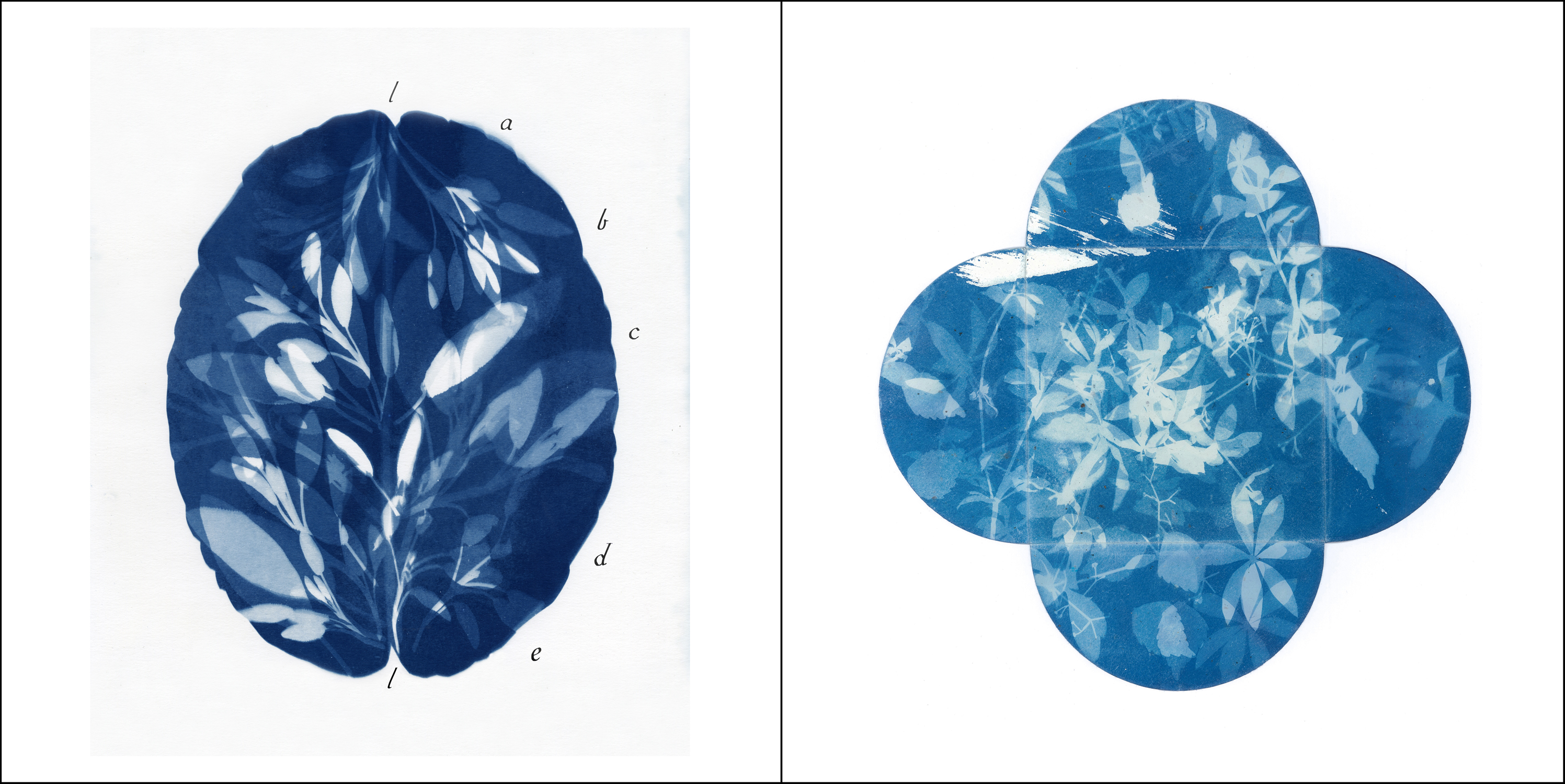
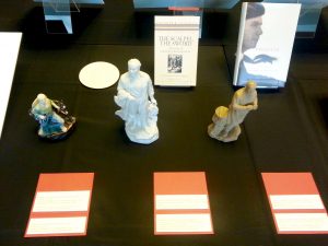
Materia Medica is an exhibition of recent work by Montreal artist Loren Williams. Invited by the Osler Library of the History of Medicine to create a body of work on the theme of Montreal’s medical history, and recipient of the Michele Larose – Osler Library Artist-in-Residence Programme for 2017, the artist combines artifacts from the Osler collection with collected and created traces of Montreal’s medical past.

The work in this exhibition draws inspiration from books and artifacts in the Osler Library as well as a wide variety of other sources. In particular, early maps of the city offer a form of time travel, indicating the location of the first hospitals and their large gardens used for food and medicinal plants. Three hundred year old streets such as rue de l’Hopital and rue des Soeurs Grises still exist in Montreal today, drawing direct lines to Montreal’s medical history, as do streets named Jeanne Mance, Marguerite d’Youville and Penfield.

Interested in these traces that reference Montreal’s medical heritage, Loren Williams has created a body of images that form a shadow archive. Images of medicinal plants used by the First Peoples and early settlers in Montreal were created using a 19th century camera-less photographic process called Cyanotype. The blue coloured images reveal the shadowy forms and details of the plants. They are like paper X-rays, made from a simple photographic process that uses UV rays, ie. sunlight, to expose the image, and water to develop it, sun and water­­ being the same basic ingredients required by plants.

Over the course of a year, the artist followed charted streets and routes that link Montreal’s past and present. Using epidemiological maps, she explored the sites and neighbourhoods of the city’s devastating outbreaks of Typhus, Cholera, Small Pox and Tuberculosis. Other plans of the city led her to sites of hospitals, asylums and the longest duel in Canadian history over the building of a new hospital.

Like the collected plants that echo an early botanical pharmacy, Loren Williams also collected and created other traces of medical history. X-rays and teeth molds reveal the body’s structures, fractures and medical interventions. First aid kits and their compartments double as garden plans for medicinal plants, while hospital architecture is represented in the form of postcards the shape of library index cards.

These works, presented with artifacts from the Osler Library collection, bring together images and objects from the realm of science, art and everyday life, offering an eclectic, less rational, interconnected perspective of Montreal’s medical history.

—–

We acknowledge the support of the Canada Council for the Arts, which last year invested $153 million to bring the arts to Canadians throughout the country.
Nous remercions le Conseil des arts du Canada de son soutien. L’an dernier, le Conseil a investi 153 millions de dollars pour mettre de l’art dans la vie des Canadiennes et des Canadiens de tout le pays.


Loren Williams is a visual artist working predominantly in photography. The passage of time, natural history, museums and obsolete photographic technologies are the inspiration and vocabulary of her practice. Frequently there is a connection between the artwork and the site where it is presented.

Originally from the Kootenays in British Columbia, Loren Williams moved to Montreal in 1993 and received her BFA honours in photography at Concordia University. She has received awards and grants from the federal and provincial art councils and has exhibited her work nationally.

Loren Williams extends much gratitude to Michele Larose, the Osler Library for the History of Medicine, and the Canada Council for the Arts for their generous contribution to this residency and exhibition project.

Vernissage for a new exhibition by 2016 Larose-Osler-Artist-in-Residence Dr. Lucy Lyons, Impossible Pathologies: Re-fragmenting the Archive

Thursday, October 12, 2017, 17:30-19:00. Please come to a vernissage at the Osler Library to celebrate the opening of our newest exhibit.

RSVP osler.library@mcgill.ca

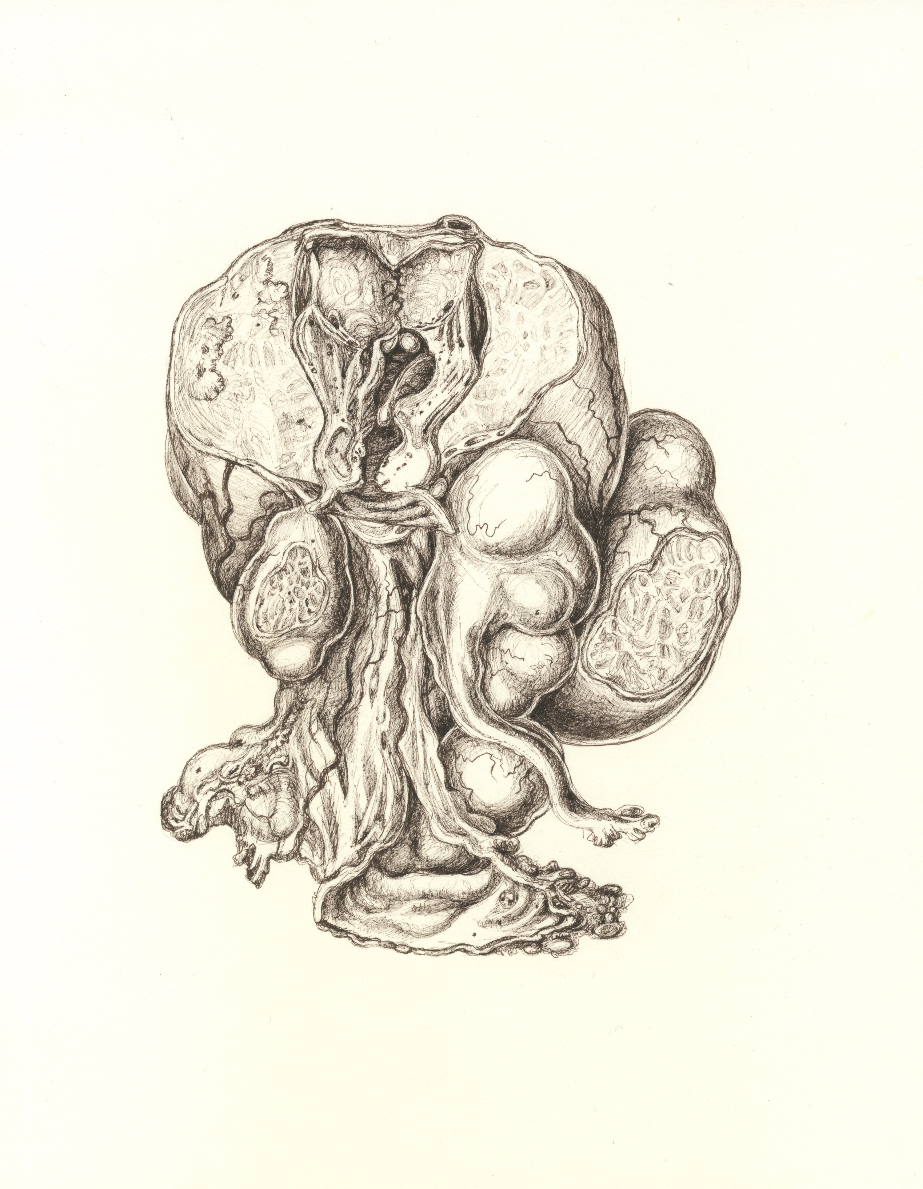
During the 2016-2017 academic year, Dr. Lucy Lyons spent time in the Osler Library archives studying the illustrations made by the English physician and medical writer, Robert Hooper. Inspired by Hooper’s method of cutting out parts of his drawings like the analogue version of Photoshop, Lyons created her own fantastic collaged composites. This composite method was then transferred into studies of the collections in the Maude Abbott Medical Museum to create new, impossible pathologies. This exhibition is an exploration of the beauty of the fragment which is synonymous with pathology. If pathology is the fractured, broken, diseased, deformed fragment of the human body, this work explores the further fracturing, breaking and then re-assembling of parts.

Dr. Lucy Lyons received her PhD from Sheffield Hallam University. Her practice focuses on drawing within medical museums and working collaboratively to explore the beauty of collections. She is especially interested in the hidden, the overlooked, insignificant or in-between. This residency will allow her to push and explore her own practice and develop work in new ways whilst bringing new audiences to the collections.

The Michele Larose – Osler Library Artist-in-Residence award, is given annually to one or more deserving candidates with a degree in Studio Arts or a related field and/or a history of exhibiting artistic work in professional venues.

Exhibition | The Gendered Cultures of Beer and Cheese: the Regulation of Human and Microbial Bodies on the Home and Industrial Scales, 1616–2017

McIntyre Medical Building Osler Library of the History of Medicine, 3rd floor, 3655 promenade Sir William Osler, Montreal, QC, H3G 1Y6, CA

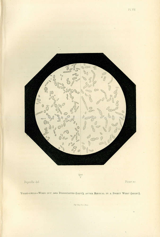
Illustration showing what healthy and worn-out yeast cells look like when viewed through a microscope. Illustration from Pasteur’s Études sur la Bière (Studies on Beer), 1876.

Vernissage: Friday, September 29, 6–8 pm at the Osler Library of the History of Medicine.

Welcome to “The Gendered Cultures of Beer and Cheese: the Regulation of Human and Microbial Bodies on the Home and Industrial Scales.” This exhibition, comprised of medical texts, cookbooks, training manuals, and industry documents, showcases the ways in which advice about best fermentation practices has changed over time. As you visit the exhibition, we hope you will consider the following questions: How is the language employed around ideas of public health, food, and alcohol production gendered and classed? Are ideas about “what is safe” and “what is dangerous” regarding fermentation practices restricted to scientific understanding? To what degree are these ideas socially embedded concepts?

The materials for this exhibit come from McGill University’s Osler Library of the History of Medicine; Rare Books and Special Collections; the Schulich Library of Physical Sciences, Life Sciences, and Engineering; the MacDonald Campus Library; the Blackader-Lauterman Collection of Architecture and Art; and the private collection of Alex Ketchum.

This exhibit has been made possible by the generous support of McGill University’s Faculty of Medicine; the Institute of Gender, Sexuality, and Feminist Studies; the Department of History and Classical Studies; the Department of English; and the Office of the Vice Principal’s Research and Innovation Grant.

We are pleased to offer this exhibit as part of this fall’s conference, “Leavening the Conversation: Food, Feminism, and Fermentation.” The event will be taking place on McGill’s campus from September 29 to October 1st. In addition to the above sponsors, the larger event is also sponsored by Le Réseau québécois en études féministes of L’Université du Québec à Montréal, Concordia University’s Department of Sociology and Anthropology, Concordia University’s Centre for Sensory Studies, and the University of Alabama’s Department of Gender and Race Studies.

For more information, please visit: foodfeminismfermentation.com

Curated by Alex Ketchum, PhD candidate, Department of History and Classical Studies

For Osler Library of the History of Medicine’s opening hours, please click here.

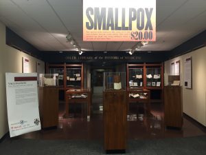
Montreal and the History of Vaccination Debates at the Osler Library

La version française suit

“The recent smallpox epidemic in Montreal – vaccinating American-bound passengers on a train of the Grand Trunk Railway,” by Marvin James, photo-mechanical reproduction of a wood engraving (1885). Osler Library Prints Collection, OPF000001.

Montreal 375 gives us the opportunity to reflect on the parcours of our city, to explore its past, and to bring this history into dialogue with our present. Our current exhibit at the Osler Library not only highlights the richness of our collections, but also brings them into contemporary conversations in a very Oslerian way. Sir William Osler collected his monumental library of the history of medicine not simply for the intellectual content or the beauty of the books and objects, but because he believed that we live with the past everyday: “the past,” Osler writes in his famous speech, Aequanimitas, “is always with us; never to be escaped; it alone is enduring.”

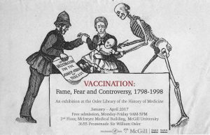
Our newest exhibition, “Vaccination: Fame, Fear and Controversy, 1798-1998,” engages with local examples of historical vaccine controversies in an attempt to understand the enduring fear and hesitancy surrounding vaccination. Curators Dr. Rob Boddice of the Freie Universitat Berlin and Cynthia Tang, PhD candidate in the Department of Social Studies of Medicine/Department of History and Classical Studies, showcase the arguments made by pro- and anti-vaccinationists during the 200 years following Edward Jenner’s invention of vaccination.

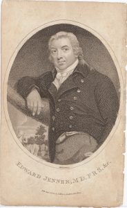
Stipple engraving of Edward Jenner by Mackenzie (London: T. Hurst, 1802). Osler Library Prints Collection, OP000643.

In 1798, Jenner, an English country physician, published the results of his experimental use of matter from cowpox pustules to inoculate patients against smallpox, which has long been endemic to industrialized cities like Montreal. This procedure, which Jenner named vaccination, was swiftly implemented as a standard public health measure and vials of vaccine were shipped and carried worldwide. However, vaccination’s benefits and promises were attended by controversy from its earliest days. Already at the beginning of the 19th century, popular pamphlets and images stoked public anxieties about the introduction of animal material and disease agents into the body. Even after the World Health Organization proclaimed smallpox eradicated in 1980, the debates surrounding vaccination that began in the 19th century have persisted in our public discourse.

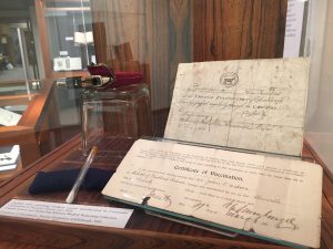
Glass capillary tube containing smallpox vaccine (Toronto, ca. 1939). Canada Science and Technology Museum, Medical Technology, art. no. 2002.0101.

A public panel presentation connected to the exhibition invited McGill and Montreal community members to think about how we understand such past examples of vaccine hesitancy and connect them to modern concerns and mythologies around vaccination. The two co-curators were joined by scholars Professor Andrea Kitta (East Carolina University), a folklorist specializing in medicine, belief and the supernatural, with a particular focus on vaccination, and late Dr. Mark Wainberg (McGill University), director of the McGill University AIDS Centre at the Montreal Jewish General Hospital, and outspoken critic of anti-vaccination rhetoric. The evening’s panel providing a wide-ranging and multidisciplinary perspective. More evidence as to the cross-institutional partnerships developed for this exhibition and the event is the blog post, “Hope and Fear in a Glass Capillary: Connecting over the History of Medicine with the Osler Library,” written by curator Cynthia Tang for the Canada Science and Technology Museum blog. In it, she explores a single artifact: a glass capillary tube containing smallpox vaccine manufactured Toronto circa 1939, and all of the hopes and fears contained within.

We are pleased to announce that this exhibition’s run has been extended through the summer! All are welcome to come view the exhibition during library opening hours (Monday through Friday, 9:00-5:00) in the Osler Library, 3rd floor, McIntyre Medical Building, 3655 Promenade Sir-William-Osler.

Montréal et l’histoire des débats sur la vaccination à la bibliothèque Osler

« L’épidémie récente de variole à Montréal — la vaccination des passagers américains dans un train du Grand Trunk Railway », par Marvin James, reproduction photomécanique d’une gravure sur bois (1885). Collection d’images de la bibliothèque Osler, OPF000001.

Le 375e anniversaire de Montréal nous donne l’occasion de nous replonger dans le parcours de notre ville, d’explorer son passé et d’en discuter dans le contexte actuel. En plus de mettre en évidence la richesse de nos collections, la nouvelle exposition présentée à la bibliothèque Osler permet d’engager des conversations les concernant à la manière d’Osler lui-même. Sir William Osler a construit cette riche bibliothèque sur l’histoire de la médecine, non seulement pour l’héritage intellectuel ou la beauté des livres et des objets qui s’y trouvent, mais aussi parce qu’il croyait que le passé fait partie de notre vie de tous les jours. « [traduction] Le passé, écrivait Osler dans son célèbre discours, Aequanimitas, fait toujours partie de nous et nous ne pouvons y échapper; c’est le seul qui résiste à l’épreuve du temps. »

Notre nouvelle exposition, « Vaccination : Fame, Fear and Controversy, 1798-1998 », offre des exemples de controverses autour de la vaccination, qui faisaient rage dans la région à l’époque, en vue de permettre aux visiteurs de comprendre la peur et l’hésitation qui régnaient à son égard. Les conservateurs Rob Boddice de la Freie Universitat de Berlin et Cynthia Tang, candidate au doctorat au département des Sciences sociales en médecine et au département de l’Histoire et études classiques, présentent le raisonnement des personnes qui étaient en faveur de la vaccination et de celles qui s’y opposaient au cours des 200 ans qui ont suivi la découverte de la vaccination par Edward Jenner.

Portrait d’Edward Jenner par gravure au pointillé réalisé par Mackenzie (Londres : T. Hurst, 1802). Collection d’images de la bibliothèque Osler, OP000643.

En 1798, Jenner, un médecin d’Angleterre qui pratiquait en région rurale, a publié les résultats de ses expériences sur l’utilisation de matières extraites de pustules contenant le virus de la vaccine pour inoculer les patients contre la variole, qui a longtemps été endémique dans les villes industrialisées comme Montréal. Cette méthode, que Jenner a nommée vaccination, a été rapidement adoptée et généralisée comme mesure de santé publique, et des fioles de vaccin antivariolique ont été expédiées et distribuées dans le monde entier. Toutefois, les avantages et les résultats prometteurs de la vaccination ont dès le début fait l’objet d’une controverse. Déjà, au début du 19e siècle, la publication de brochures et d’images suscitait dans la population des inquiétudes liées à l’introduction de matière animale et d’agents pathogènes dans le corps. Même après l’annonce de l’Organisation mondiale de la santé en 1980, selon laquelle la variole avait été éradiquée, les débats publics amorcés au 19e siècle autour de la vaccination se sont poursuivis.

Tube capillaire contenant le vaccin antivariolique (Toronto, vers 1939). Technologie médicale, Musée des sciences et de la technologie du Canada, nº d’artefact : 2002.0101.

Dans le cadre de l’exposition, une présentation publique a été dirigée par un groupe d’experts à laquelle ont été conviés les membres de la collectivité de McGill et de Montréal. Cette initiative a porté les participants à réfléchir sur ces exemples passés de l’hésitation autour de la vaccination et à faire le lien avec les préoccupations et mythes sur la vaccination qui perdurent encore aujourd’hui. Les deux conservateurs étaient accompagnés des chercheurs Andrea Kitta (East Carolina University), une folkloriste spécialiste de la médecine, des croyances et du surnaturel, qui s’intéresse particulièrement à la vaccination, et feu Mark Wainberg  (Université McGill), directeur du Centre universitaire sur le SIDA de l’Université McGill à l’Hôpital général juif de Montréal et critique de la rhétorique anti-vaccin. La présentation tenue en soirée a permis de dégager une perspective générale et multidisciplinaire. Le billet de blogue « Hope and Fear in a Glass Capillary: Connecting over the History of Medicine with the Osler Library » ([traduction] Espoir et peur dans une fiole de verre — Retracer l’histoire de la vaccination en compagnie de la Bibliothèque Osler), publié par la conservatrice Cynthia Tang pour le Musée des sciences et de la technologie du Canada, témoigne des partenariats interorganisationnels établis pour l’exposition et l’événement. Son bloque porte sur un seul objet : une fiole de verre contenant le vaccin contre la variole fabriqué à Toronto vers 1939, et tous les espoirs et les peurs qu’elle suscitait.

Nous sommes heureux d’annoncer que cette exposition se poursuivra durant l’été! Vous êtes tous invités à venir voir l’exposition durant les heures d’ouverture (du lundi au vendredi de 9 h à 17 h) à la bibliothèque Osler, 3e étage de l’édifice McIntyre Medical Sciences, 3655, promenade Sir-William-Osler.


A Collaborative Exhibit for the Chinese Premier

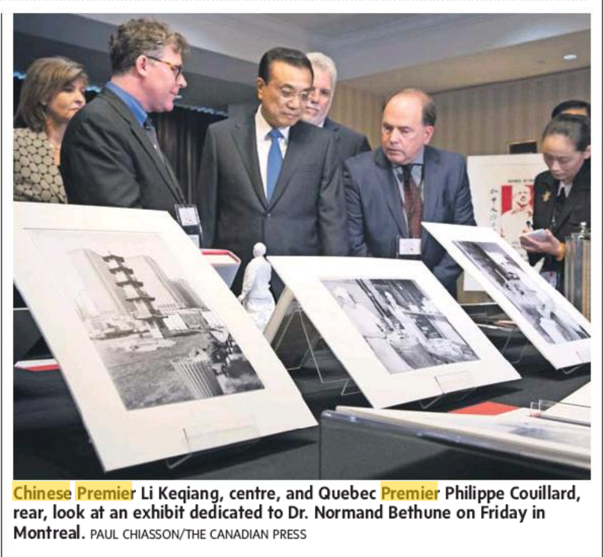
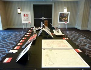
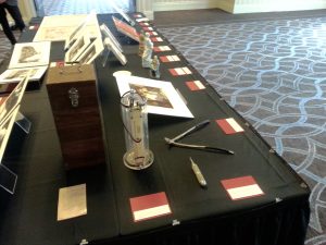
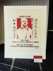
Last month it was our pleasure to work in collaboration with McCord Museum and Bibliothèque et Archives nationales du Québec (BAnQ) on a private exhibition for Chinese Premier Li Keqiang during his visit to Montreal. The combined curatorial effort resulted in a showcase of items with historical ties and strong cultural connections between Québec and China. The exhibit included maps, photographs, books, posters, and artifacts, with a particular emphasis on the great physician Norman Bethune — a McGill graduate and well-known Canadian hero in the People’s Republic of China.

Screen Shot 2016-09-27 at 10.54.10 AM

Chinese Premier Li Keqiang (centre) views the exhibit with Osler Librarian Chris Lyons (front left) and Quebec Premier Philippe Couillard (back). The Globe and Mail, Saturday Sepetember 24, 2016. Photo credit: Paul Chiasson/The Canadian Press.

We welcome and look forward to more collaborative exhibits like this one in the future!

Science Literacy Week

sci-lit-week-1200McGill campus is gearing up for Science Literacy Week 2016, happening all through next week September 19-25th. The Osler Library will host a special guided tour of Knowing Blood: Medical Observations, Fluid Meanings with curators Darren N. Wagner and Nick Whitfield on Monday, Septemebr 19th @ 11:30am. Registration is not required, but feel free to sign up to let us know you are coming.

For more details and a full listing of next week’s events click here!

New exhibition: Knowing blood / Sang sens

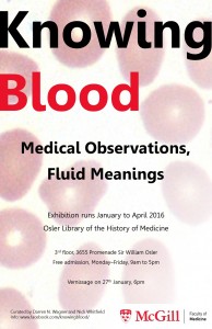
Knowing BloodKnowing Blood: Medical Observations, Fluid Meanings

This exhibition is about observations, meanings, and understandings of blood from the late 15th to the mid-20th century. Blood has long been a powerful and evocative symbol, signifying themes from life, identity, community and kinship to sex, lineage, violence and death. Practices of observing blood in experiment, diagnosis, and therapy have also varied widely: a melange of cells seen under a microscope, a pulse felt by a trained touch, the taste of blood from a barber-surgeon’s bowl, a map comparing hematological and racial groups. Modern Western medicine has known not one but many kinds of blood.

Knowing Blood queries how various practices of observation have encountered the multifarious meanings of blood and negotiated new medical knowledge. The objects, texts, and images displayed here are drawn from the collections of the Osler Library of the History of Medicine, the Humanities and Social Sciences and Schulich Libraries, the Maude Abbott Medical Museum, and le Musée des Hospitalières de l’Hôtel-Dieu de Montréal. Five thematic cases highlight different historical approaches to observation, their relation to changing systems of medical practice and to blood’s broader meanings. We invite you to explore this rich 400-year history of knowing and observing the most vital of bodily fluids.

Sang sensSang sens : observations médicales, interprétations fluides

Cette exposition porte sur l’observation, la signification et les représentations du sang de la fin du XVe au milieu du XXe siècle. Évoquant des thèmes de vie, d’identité, de communauté et de parenté, mais aussi de sexe, de génétique, de violence et de mort, le sang constitue depuis des siècles un symbole puissant. Les pratiques d’observation du sang dans les contextes expérimental, diagnostic et thérapeutique ont par ailleurs largement varié; depuis un mélange de cellules aperçu sous un microscope à un pouls tâté minutieusement, en passant par une certaine odeur détectée après une saignée ou encore une carte du monde comparant divers groupes hématologiques et raciaux. La médecine occidentale contemporaine a ainsi connu non pas un mais bien plusieurs types de sang.

Sang sens interroge les croisements multiples entre ces diverses pratiques d’observation et d’interprétation ayant contribué à la mise en place de nouveaux savoirs médicaux. Les objets, textes et images ici exposés sont tirés des collections de la bibliothèque Osler d’histoire de la médecine, de la bibliothèque des sciences humaines et sociales et de la bibliothèque Schulich, du Musée médical Maude Abbott, et du Musée des Hospitalières de l’Hôtel-Dieu de Montréal. Cinq vitrines thématiques mettent en évidence différentes facettes historiques de l’observation ainsi que leur relation avec les systèmes de la pratique médicale et les significations variées du sang dans divers discours. Nous vous invitons à découvrir cette histoire riche de 400 ans au sein de laquelle a été imaginé et montré le plus vital des fluides corporels.


The vernissage will be held on January 27th at 6PM and is open to all / Le vernissage aura lieu le 27 janvier à 18h et est ouvert a tous.

The exhibition is accessible during library opening hours, Monday-Friday, 9AM-5PM/ L’exposition est accessible pendant les heures d’ouverture de la bibliothèque, lundi à vendredi, 9h-17h.

Osler Library of the History of Medicine, McGill University, McIntyre Medical Building, 3655 Promenade Sir-William-Osler 


History of biology in the Osler Library

The history of natural history and biology is heavily represented in the collection of the Osler Library, not just because of their important place in the history of medicine, but also in the life story of the library’s founder.

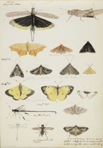
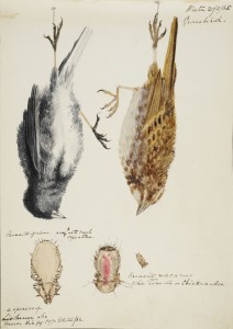
Coloured sketches by W. A. Johnson. William Arthur Johnson Fonds, P139, Osler Library

Coloured sketches by W. A. Johnson.
William Arthur Johnson Fonds, P139, Osler Library

William Osler (1849-1919) grew up in Bond Head, Ontario, surrounded by books. His father’s theological library had around 1,500 items. The young Osler had his first glimpse of a scientific library when he left home to attend Trinity College School (located today in Port Hope, Ontario). The Warden of the school, Reverend W.A. Johnson (1816-1889), according to Osler, “a good friend, botanist, a practical palaeontologist, an ardent microscopist,” inspired Osler with a passion for natural history.

While studying Anglican divinity at Trinity College, Toronto, Osler spent two years living with James Bovell, the Chair of Natural Theology at Trinity College and a member of the Toronto School of Medicine faculty, assisting him in his work. He would often go out to collect various samples of algae and other specimens to fix onto slides for Bovell. His first publication, “Christmas and the Microscope” (1869) demonstrated his love for and proficiency at microscopy. Osler transferred from Trinity College to the Toronto School of Medicine, and then ultimately to the Medical Faculty at McGill University to pursue the greater clinical opportunities available to medical students in Montreal.

Coloured sketches by W. A. Johnson. William Arthur Johnson Fonds, P139, Osler Library

Coloured sketches by W. A. Johnson.
William Arthur Johnson Fonds, P139, Osler Library

Osler’s affinity for natural history flourished during his stay at McGill. Through another mentor, Dr. Palmer Howard, Dean of the McGill Faculty of Medicine, Sir William was introduced to some of the foundational works in the field of life sciences, including those of Laennec, Stokes, and Graves. Osler’s thesis was based on the preparation of gross and microscopic slides from twenty autopsies. After graduation, he considered the India Medical Service, but opted to remain in Montreal, so as to pursue his work in microscopy. He was offered the Chair of Botany at McGill because of this expertise, but turned the position down in favour of a lectureship in Physiology. He became Chair of Clinical Medicine at the University of Pennsylvania in 1884, where he was a member of the Biology Club. His bibliophilia was inspired by places such as the College of Physicians in Philadelphia. Osler moved to the Johns Hopkins University in 1889, where in 1893 he played an instrumental role in the creation of the Johns Hopkins Medical School and teaching hospital.  He was appointed Regius Professor of Medicine at Oxford University in 1905. At Oxford he served equally as a curator of the Bodleian Library and was heavily involved in the work of the Bibliographical Society of London. In Oxford, he had the leisure time to devote himself to his passion for book collecting, designing a library that would contain the most significant works produced in the history of medicine and science.

Come see a selection of works of natural history and biology in a special exhibition up now in the Osler Room of the Osler Library.


On the Surface/Skin deep: Interview with the curators

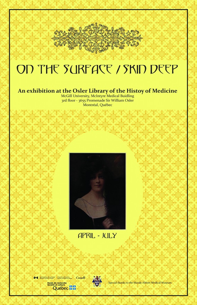
On the Surface Skin Deep posterSylie Boisjoli and Shana Cooperstein are doctoral students in art history in the Department of Art History and Communication Studies at McGill. They are the guest curators of our current exhibition, On the Surface/Skin Deep. They agreed to answer a couple questions about the exhibition and their work.

AD: First, could you each tell me a little bit about your research interests, and current work?

SB: I’m currently working on my PhD thesis in the Department of Art History and Communication Studies and am the course supervisor for the Museum Internship class, which aims to accompany and enhance students’ internships at museums or galleries. During my MA in art history, which I also did at McGill, I conducted extensive primary research at the Osler Library for my major research paper on late nineteenth-century French artists’ visualisations of the development of serum therapy treatments against diphtheria. My PhD work has now shifted to investigate the emergence of the notion of prehistoric time in France within wider nineteenth-century scientific and philosophical debates around Darwinian evolutionary theories on the progress and mutability of the human species. By examining the social, political, and intellectual conditions in which the idea of prehistory developed, I analyze how representations of prehistoric people and places normalized the belief that time was a dynamic force that could drive forward or limit France’s progress as a nation. My work thus explores the increased public interest in representations and conceptions of prehistoric time in relation to the wider nineteenth-century impulse for reading the past and the passage of time in/on the body. My dissertation also considers the notion of prehistory as an indicator of, and participant in, a significant shift in how French society understood its national history. The fascination with prehistory was certainly not restricted to a few specialists in the human sciences. Instead, my research investigates ways in which artists, writers, and scientists disseminated their visions of the past across the French public sphere.

SC: After receiving a Master of Arts in art history at Temple University, I began pursuing a doctorate in art history at McGill University. My personal trajectory has led me to develop an interest in theories of representation, standardization, and the intersection of art and science. Bridging these areas of inquiry, my dissertation situates nineteenth-century French drawing pedagogy at the nexus of art, industrial design and the theories of knowledge. Specifically, I explore the institutionalization and reorganization of drawing in public pedagogical programs, design schools, and at the École des Beaux-Arts. In the past, I have had the opportunity to intern at various institutions, such as The Institute of Contemporary Art in Philadelphia, The Museum of Modern Art, and The Carnegie Museum of Art. I also have authored an article in Leonardo titled: “Imagery and Astronomy: The Visual Antecedents Informing Non-Reproductive Depictions of the Orion Nebula.”

AD: What led you to choose the topic of the exhibition? How did you go about exploring the topic and selecting materials?

SB: One of the most striking images in the Osler Library’s collection is the “The Flayed Angel” from an eighteenth-century medical atlas by French anatomist and printmaker Jacques Fabien Gautier D’Agoty (c.1716-1785). In this richly coloured mezzotint, a woman’s back skin is peeled away to reveal the inner anatomy of her spine and the musculature of her back. The idea of slicing and paring a portion of the body’s outer boundary led us to question how skin has been conceived as the interface between the body’s depths and surface. Although D’Agoty’s fascinating picture was at the starting point of our thinking process, eighteenth-century mezzotints and prints will (unfortunately!) have to wait for another exhibition. As specialists in nineteenth-century French art and because we knew we could display wonderful wax moulages of skin diseases from the Maude Abbott Medical Museum, we wanted to work within the scope of that century. Moreover, it’s a time when dermatology emerged as a specialized branch of medicine concerned with visualizing and diagnosing skin. Once we began to explore the vast and varied nineteenth-century materials on skin from the Osler collection, it became clear that images had played a significant role in disseminating ideas on what doctors and beauty specialists considered to be normal and healthy skin ―and thus how to diagnose skin diseases and other epidermal conditions. Indeed the authors of many of the medical texts on display in the exhibition stated that medical images were indispensable to teaching doctors how to correctly scrutinize the surface of the body. I was especially interested in why several doctors included the superficial alteration of skin pigmentation in their treatises on skin diseases. In Élie Chatelain’s Précis iconographique des maladies de la peau from 1893, the doctor provided little in the way of an explanation for the cause of vitiligo, and even less for its treatment. Yet Chatelain included a hand-painted photograph that highlights the difference between a male patient’s skin colour and patches of depleted pigment that emerged on his arms, shoulders, torso, and groin. Because of, and due to, the emphasis put on the visible aspects of skin diseases, we hope that visitors at the Osler Library will see how images were part of a medical discourse that considered changes in skin colour, for example, to be a pathological feature. By examining the medical images on display, we hope to raise larger questions about the place of medical discourse in debates about race, class, and gender in nineteenth-century society.

SC: Originally, the exhibition was scheduled to open a year ago to coincide with a guest lecture by Dr. Mechthild Fend, an art historian interested in eighteenth- and nineteenth-century representations of the body, titled “Body to Body: The Dermatological Wax Moulage as Indexical Image.” Although we were unable to move forward with the project until the Osler reopened this year, Dr. Hunter encouraged us to build from Dr. Fend’s scholarship. In doing so, we explored how art historians can profit from medical collections by analyzing depictions of skin from a humanistic perspective.
First and foremost, the availability of material at the Osler guided the scope and trajectory of the exhibition. After agreeing on the representation of epidermis in modernity as the unifying thematic of the display, I began to think about how artists depicted skin. Rather than represent individual abnormalities or eccentricities, artists historically idealized features. With this in mind, I began consulting medical atlases, scientific texts, and cosmetic handbooks with an eye toward issues of beauty and idealization, aging, and birthmarks. Tying together these themes was the ways in which abstract notions of comeliness and what constitutes comeliness shaped the representational strategies used to display “superficial” medical features. I was curious about how physicians attempted to rationalize imperfections, such as wrinkles and birthmarks, as well as how scientific “facts” lent supposedly scientific support to cosmetic “knowledge.” Likewise, I was interested in how the rationalization of imperfection often defied the well-known expression: “beauty is only skin deep.” For instance, the physiognomic study of wrinkles connected the appearance of skin to the deepest recesses of the mind.

AD: Could you describe your curatorial vision or approach to the exhibition? What have you hoped to achieve?

SB: As art historians, a consideration of how skin has been depicted in paintings that are then reproduced in beauty manuals, how skin and the diseases, blemishes, or creases that can mark it have been moulded into wax, photographed or drawn, led us to question how skin has been historically conceived. This is also why we chose to look at a diverse range of materials: treatises on hygiene, beauty manuals, as well as atlases and guides for professional medical practitioners. We believe that this allows us to explore the wider historical and social contexts in which medical knowledge on skin developed. We also hope that visitors will be able to explore the historical roots for the ongoing pressure and pursuit to have smooth and evenly pigmented skin.

SC: As previously mentioned, the broader aim of this exhibition was to convey how art historians can use medical collections. Personally, I hoped to convey the importance of interdisciplinary research to an audience with diverse backgrounds without compromising the complexity of academic scholarship. Several art historians use interdisciplinary research to highlight how other disciplines, such as medicine, shaped artistic production. I sought–and in my dissertation will continue–to do the reverse. I aim to establish how art shapes the way other disciplines have understood and continue to understand their practice. In the case of early dermatology, the most obvious manifestation of this is that physicians often hired illustrators trained in the fine arts and with particular conventions to represent case studies.

AD: What sorts of other exhibition experiences have you had or been involved in? Have you found there to be particular considerations or challenges involved in working with rare books?

SB: During my undergraduate degree in History at the University of Winnipeg I had the opportunity to intern as a curatorial assistant at the Buhler Gallery, a unique gallery located in the Saint-Boniface Hospital that aims to provide a restful and contemplative space for visitors. While interning there I helped to curate an exhibition of two Winnipeg painters, Arthur Adamson and George Swinton, entitled Marking the Message. Before starting my MA at McGill I also worked as an Art Handler Technician at the Winnipeg Art Gallery. As far as organizing and installing exhibitions, I’m used to dealing with paintings and sculptures in conventional gallery spaces. Installing the exhibition at the Osler Library presented the special challenge of how to best display the images in delicate books. On the one hand, I’m used to handling the material at the Osler Library because of extensive research on a variety of nineteenth-century topics such as serum therapy, vaccination, diphtheria, rabies, breast feeding, and theories of degeneration. For an exhibition, however, we needed to consider how to best keep the books safely open for the duration of the exhibition. This is where the expertise of the staff was a necessary part of the process (thank you Anna, Bozena, Lily, and Chris!).

SC: I have been privileged to conduct research as an intern at several museums including The Museum of Modern Art, the Institute of Contemporary Art in Philadelphia, the Carnegie Museum of Art, the University Art Gallery at the University of Pittsburgh, and at the American Jewish Museum of Pittsburgh. These experiences offered invaluable insight into the nature of curatorial research and exhibition planning, as well as how to handle art objects. Building from my work as an intern, I’ve also been privileged to curate two exhibitions at Parsons The New School for Design titled Cultural Constructs (July 15-August 1, 2013) and (Sub)contracted (July 14-July 28, 2012). Each show featured work by candidates and recent recipients of a Master of Fine Arts in photography at Tyler School of Art, Temple University. Cultural Constructs explored the representational conventions associated with cultural heritage in the contemporary moment. This show questioned the ideological assumptions determining which architectural structures, monuments, and landscapes embody or define a culture and its history. Distinct from this exhibition, (Sub)Contracted tied together the work by five artists who adopt the visual conventions associated with photography’s earliest utilitarian uses. By documenting sculptures and capturing aerial landscape photographs, for instance, their work recalls photography’s first status as a “hand-maiden” to the fine arts and sciences. Working with rare books posed new and exciting challenges for me. Because so many of the texts housed by the Osler are illustrated, the collection offers an overwhelming amount of images from which to work! Along with this, rare books pose curatorial problems in so far as they need to be exhibited in cases. Showcases qualify the physical proximity between viewers and the objects, limiting the viewer’s ability to see the images clearly. This, coupled with the fact that it was impossible to adopt exhibition practices traditionally used in art museums to highlight formal properties or details, such as changing the wall color, curators using this space are required to think creatively about how to exhibit these materials. For instance, in the section on birthmarks, the addition of a bright red cloth highlighted the intricate colors used to represent angiomas and vascular naevus. Rare books also confound the pervasive method of organizing art works chronologically in exhibitions. Because so many of the books are reprinted in different editions over the course of several years, it proved to be easiest to think about the representation of skin thematically as opposed to chronologically. Admittedly, I felt self-conscious about the curatorial process because I am a teaching assistant to a course in which students were required to examine how exhibition practices, such as the way works are organized and hung on the wall, shape the viewer’s interpretation of the show! In many respects, we tried to highlight the “look” or aesthetic of older libraries.

Thank you, Shana and Sylvie! On the Surface/Skin Deep will run through July in the Osler Library exhibition space on the 3rd floor of the McIntyre Medical Building, 3655 Promenade-Sir-William-Osler.