Digital exhibition sneak preview, part 3

Missed our 2011 exhibition “Our Friend, the Sun: Images of Light Therapeutics, 1901-1944”? Here’s a sneak preview of the digital exhibition currently under construction. You can find the full exhibition catalogue by curator Dr. Tania Anne Woloshyn here. And stay tuned for more!
John Harvey Kellogg. Light therapeutics: a practical manual of phototherapy for the student and the practitioner. 2nd ed. Battle Creek, Mich. : Modern Medicine Pub. Co., 1927.

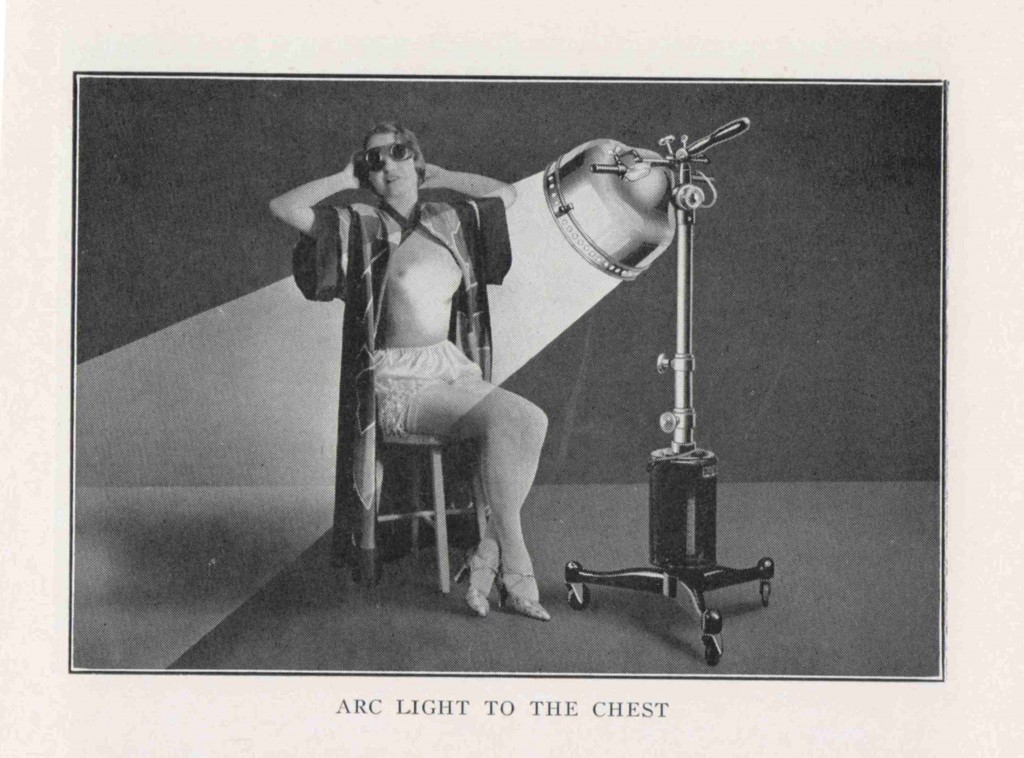
John Harvey Kellogg. Light therapeutics: a practical manual of phototherapy for the student and the practitioner. 2nd ed. Battle Creek, Mich. : Modern Medicine Pub. Co., 1927.

Remember our long-suffering patient from last week? Contrast that with this week’s gleeful recipient of light therapy. By the 1920s, physicians began incorporating advertising imagery into their textbooks. In this photomontage, we are presented with a fresh-faced, smiling model, coiffed in the latest 1920s crop. The impression is that this phototherapeutic treatment is neither uncomfortable nor distressing, but in fact an enjoyable process. The ambiguity of her surroundings, reminiscent of a photographer’s studio, is heightened by the impossibilities of the scene: the rays of the lamp (which itself appears hand drawn) shine on her chest and yet continue undisturbed beyond her.


Further reading

Simon Carter. Rise and Shine: Sunlight, Technology, and Health. New York: Berg, 2007.

Tania Anne Woloshyn, “‘Kissed by the Sun’: Tanning the Skin of the Sick with Light Therapeutics, c.1890–1930,” in Kevin Siena and Jonathan Reinarz, eds. A Medical History of Skin: Scratching the Surface (London: Pickering & Chatto, 2013), chap. 12.


Digital exhibition sneak preview, part 2

Missed our 2011 exhibition “Our Friend, the Sun: Images of Light Therapeutics, 1901-1944”? Here’s a sneak preview of the digital exhibition currently under construction. You can find the full exhibition catalogue by curator Dr. Tania Anne Woloshyn here. And stay tuned for more!

Röntgen rays and electro-therapeutics: with chapters on radium and phototherapy / by Mihran Krikor Kassabian. Philadelphia ; London: J. B. Lippincott, 1907.

Röntgen rays and electro-therapeutics: with chapters on radium and phototherapy / by Mihran Krikor Kassabian. Philadelphia ; London: J. B. Lippincott, 1907.

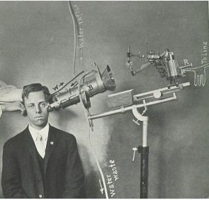
This photograph depicts a patient receiving phototherapy treatment via an arc light developed by Niels Ryberg Finsen, the Danish physician who pioneered the treatment of disease (notably lupus vulgaris) through exposure to specific wavelengths of light.

“Finsen’s method consists in concentrating actinic light, through rock-crystal lenses, on any desired part, rendered as exsanguine as possible by means of pressure, because the presence of blood acts as a barrier to the passage of the chemical rays to the tissues.” (Kassabian, 515)


Further reading

Simon Carter. Rise and Shine: Sunlight, Technology, and Health. New York: Berg, 2007.

Tania Anne Woloshyn, “‘Kissed by the Sun’: Tanning the Skin of the Sick with Light Therapeutics, c.1890–1930,” in Kevin Siena and Jonathan Reinarz, eds. A Medical History of Skin: Scratching the Surface (London: Pickering & Chatto, 2013), chap. 12.
 

Digital exhibition sneak preview

Missed our 2011 exhibition “Our Friend, the Sun: Images of Light Therapeutics, 1901-1944”? Here’s a sneak preview of the digital exhibition currently under construction. You can listen to the original exhibit talk by curator Dr. Tania Anne Woloshyn here. And stay tuned for more!

S. I. Rainforth. The stereoscopic skin clinic; an atlas of diseases of the skin, consisting of colored stereoscopic illustrations and a text in the form of clinical lectures, designed for the use of practitioners and students of medicine. New York: Medical Art Pub. Co., 1914.

S. I. Rainforth. The stereoscopic skin clinic; an atlas of diseases of the skin, consisting of colored stereoscopic illustrations and a text in the form of clinical lectures, designed for the use of practitioners and students of medicine. New York: Medical Art Pub. Co., 1914.

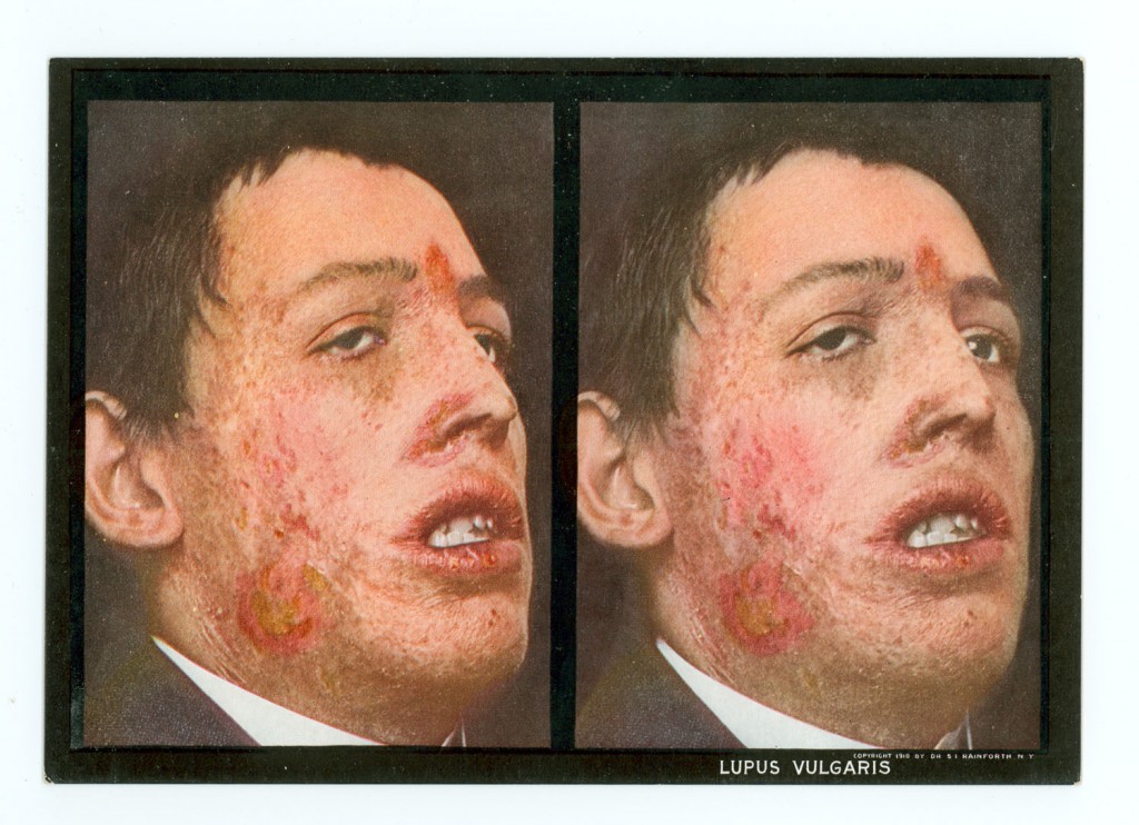
These images are designed for an early twentieth-century viewing device called a stereoscope, originally intended less for medical purposes than for entertainment. The image shows a disfiguring case of lupus vulgaris, tuberculosis of the skin, on the face of a male patient. The two photos are each shot from a slightly different angle so that when viewed together through the stereoscope they form an optical illusion in 3D.

Dr. Selden Irwin Rainforth (1879-1960) was a young physician from New York when he compiled The stereoscopic skin clinic. The work consisted of over 130 stereoscopic plates with a viewing device and was published in multiple editions. Detailed descriptions of the diseases on the back of each photo card provided useful information about skin conditions at a time when dermatologists were still rare.


Further reading

John Thorne Crissey, Lawrence Charles Parish, and K. Holubar. Historical Atlas of Dermatology and Dermatologists. Boca Raton, FL: Parthenon, 2002.

Robert Jackson. “Historical outline of attempts to classify skin diseases.” Canadian Medical Association Journal 116, no. 10 (May  1977): 1165–8. PMCID 1879511. [McGill users]

Helmut Gernsheim and Alison Gernsheim. The History of Photography: From the Earliest Use of the Camera Obscura in the Eleventh Century up to 1914. London: Oxford University Press, 1955.