Man: His Structure & Physiology

pic_2016-07-19_171814

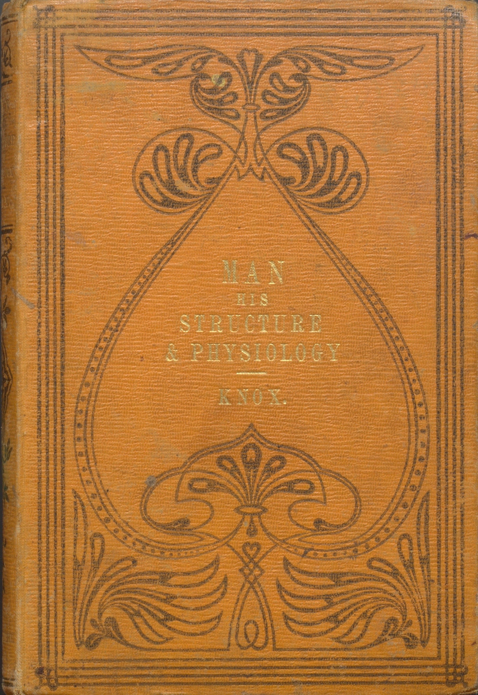
“Man: His Structure & Physiology: Popularly Explained and Demonstrated” by Robert Knox, 2nd edition, London: H. Bailliere, 1858.

This month we’ve chosen to highlight an anatomical atlas by Scottish anatomist, zoologist, and physician, Dr. Robert Knox (1791-1862). His popular book entitled Man: His Structure & Physiology: Popularly Explained and Demonstrated was originally published in 1857, with a second edition (shown here) printed a year later in 1858.

Knox was an esteemed professor at The University of Edinburgh — famous for his dissections and lectures which were often ticketed and open to the public. Prior to the 1832 Anatomy Act, it was discovered that Knox relied on illegal methods to acquire his cadavers. Knox was connected to the Burke and Hare West Port murders of 1828, and despite never being tried, his reputation was forever marred in controversy.

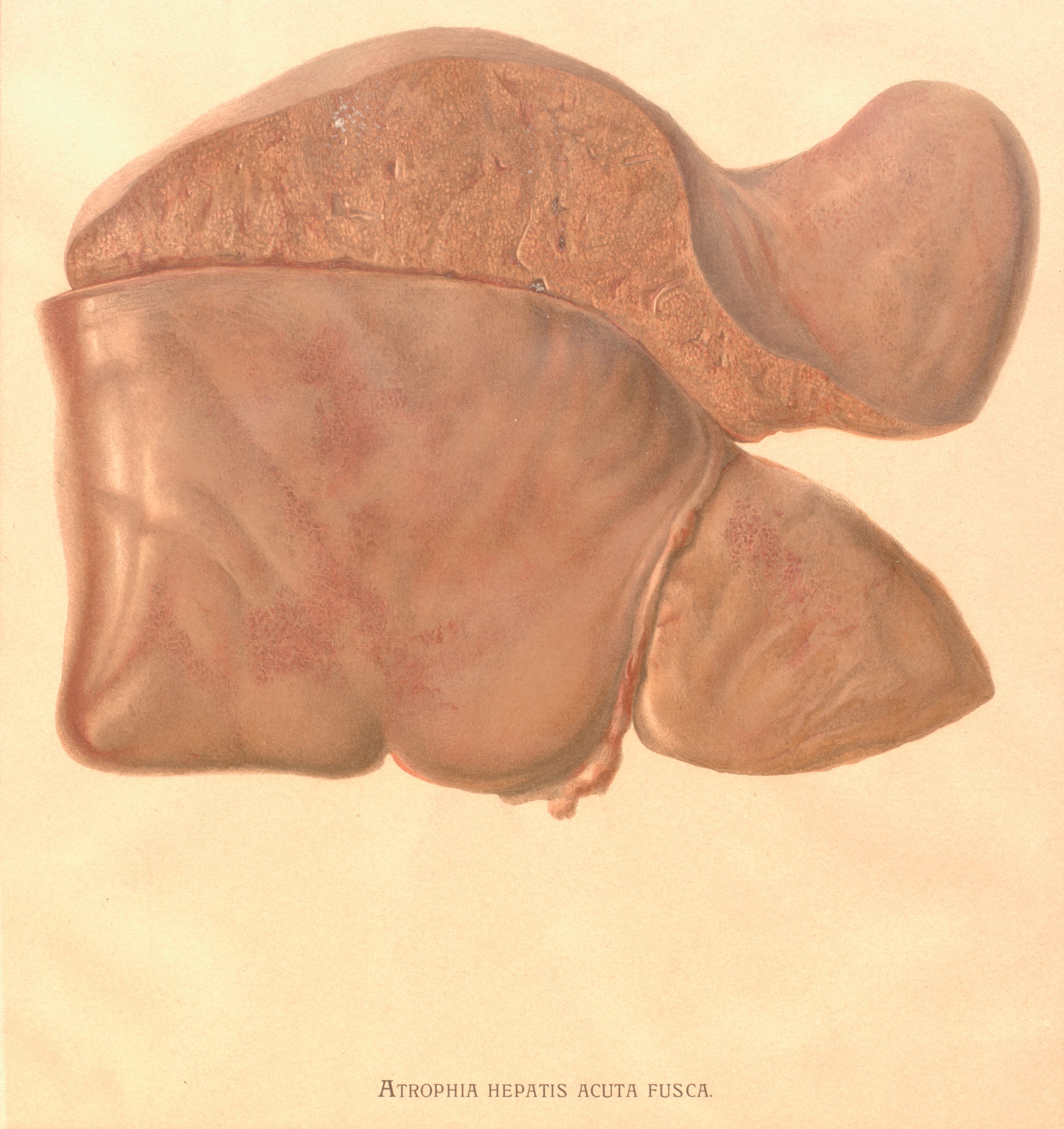
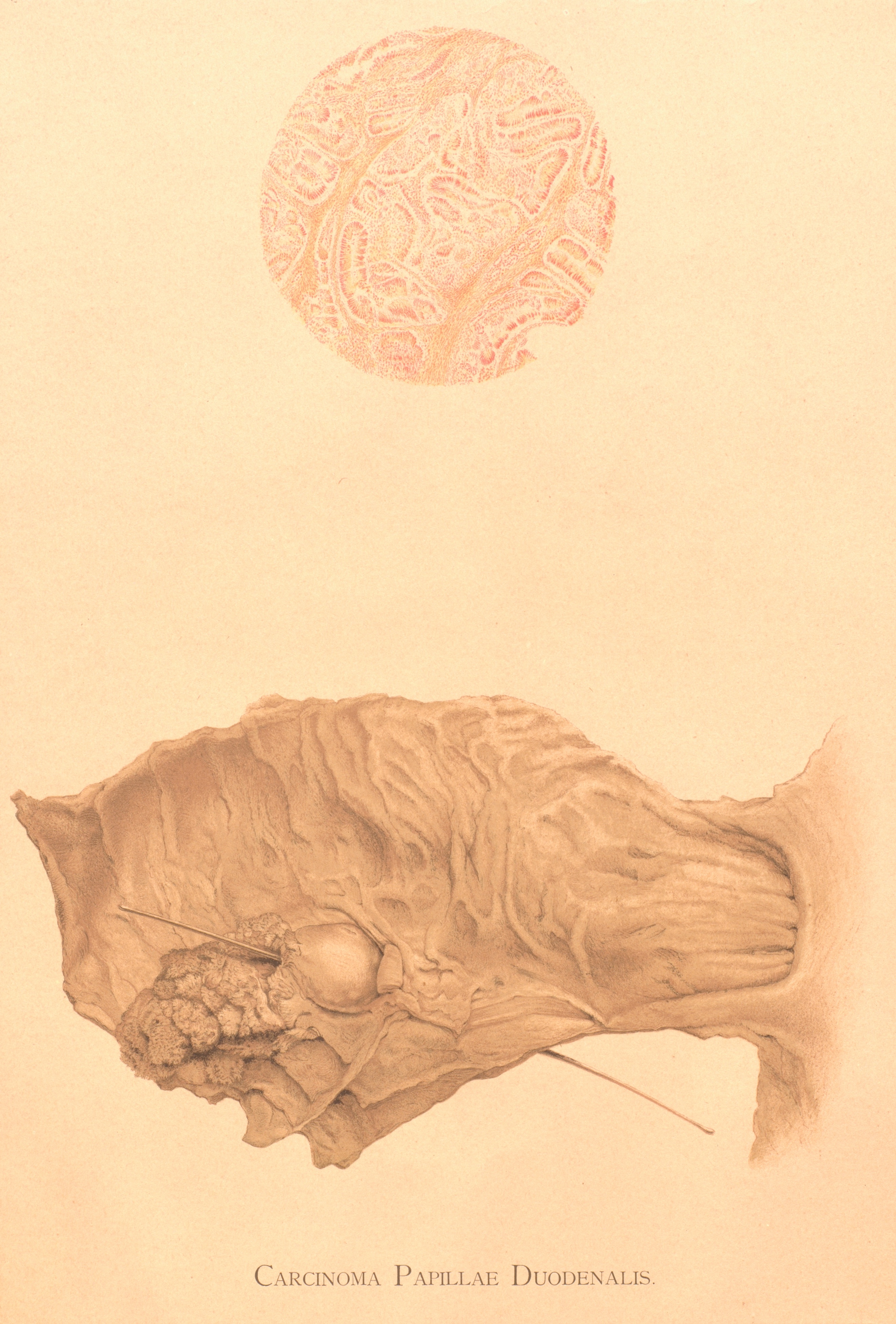
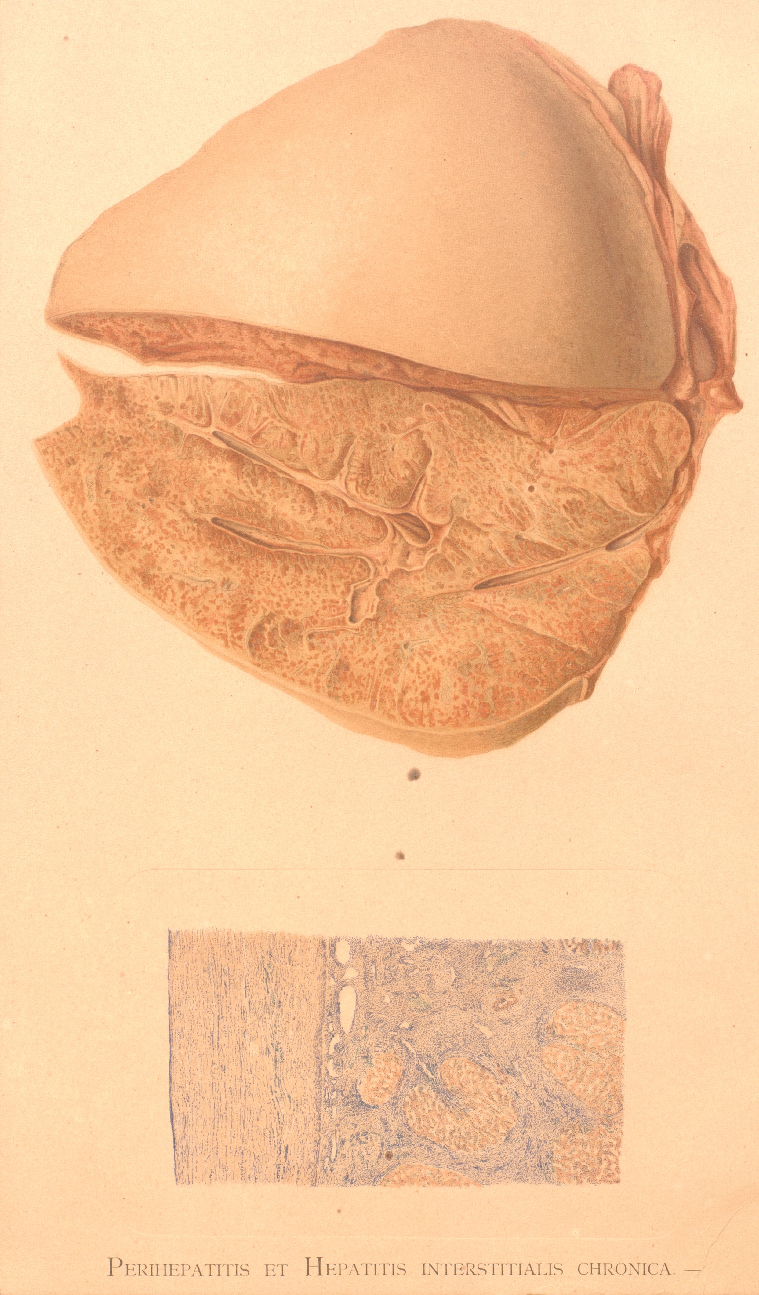
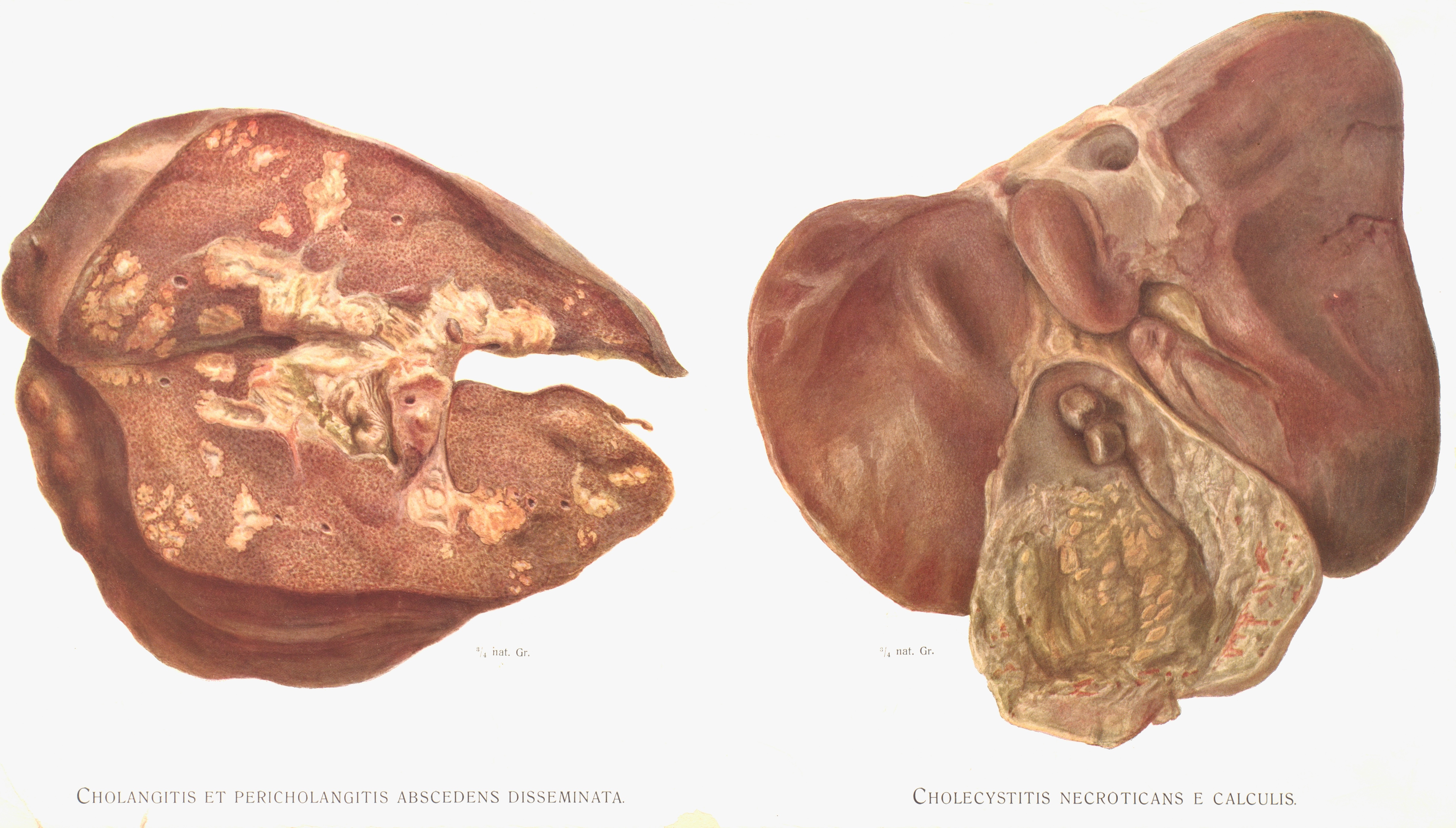
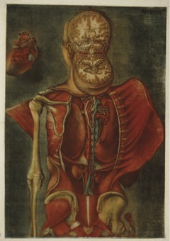
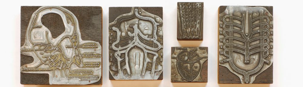
The atlas is described in simple language and includes some detailed plate illustrations — several of which can lift (“pop-up”) off the page. The idea behind this design was to imitate a dissection as much as possible, allowing students and readers to discover multiple layers of physiological detail. As the preface of the second edition describes, it is an “elementary and educational Work, containing such an outline of Human Structure and Human Physiology as may prove a safe basis whereon to build the edifice of special or philosophic inquiry and research” (London, October, 1857).

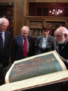
The book is available to view at the Osler Library during regular hours. For those who are not able to visit the library in person, a digitized version of a more recent pressing can be accessed at archive.org.

Version 2

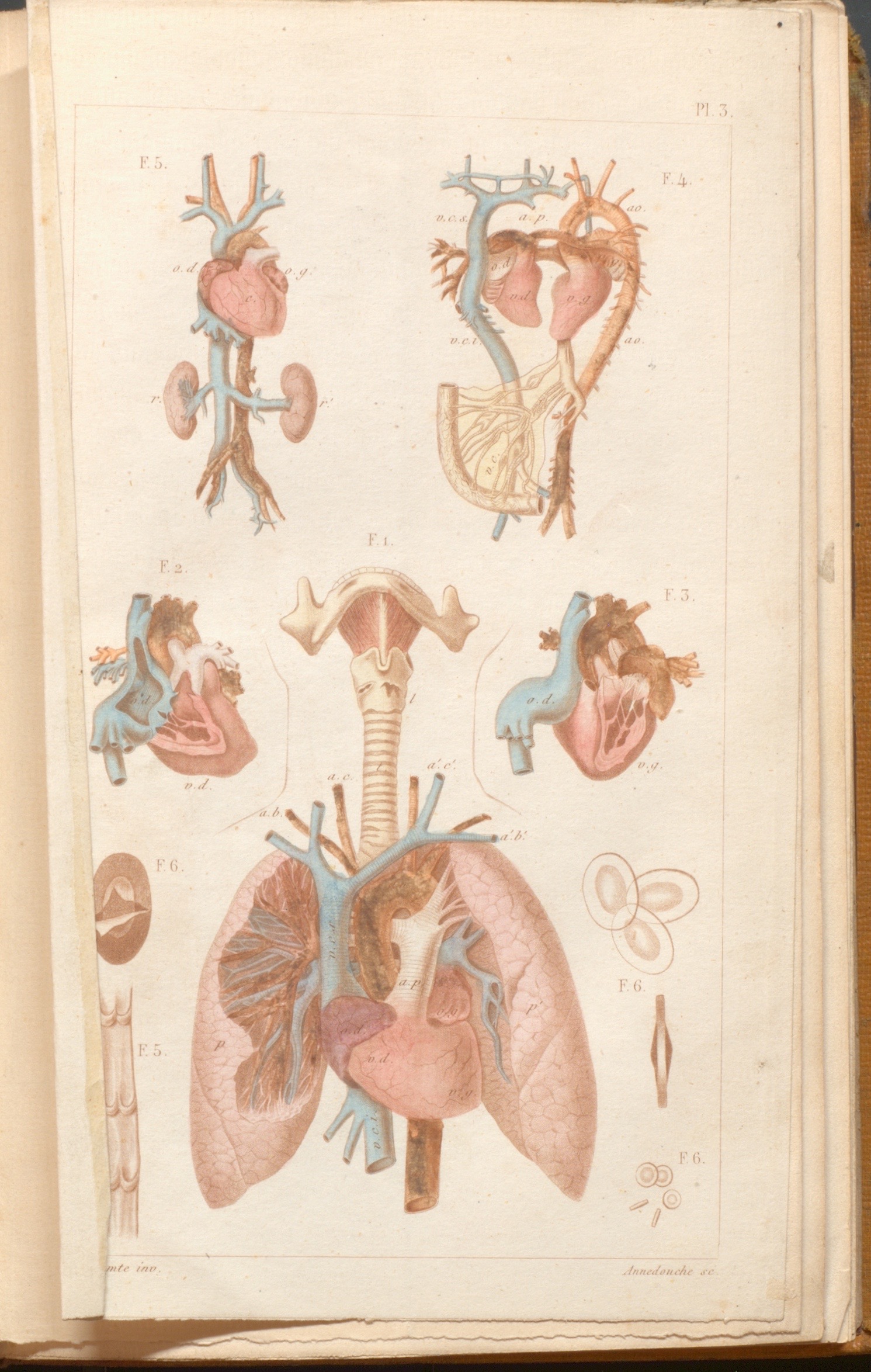
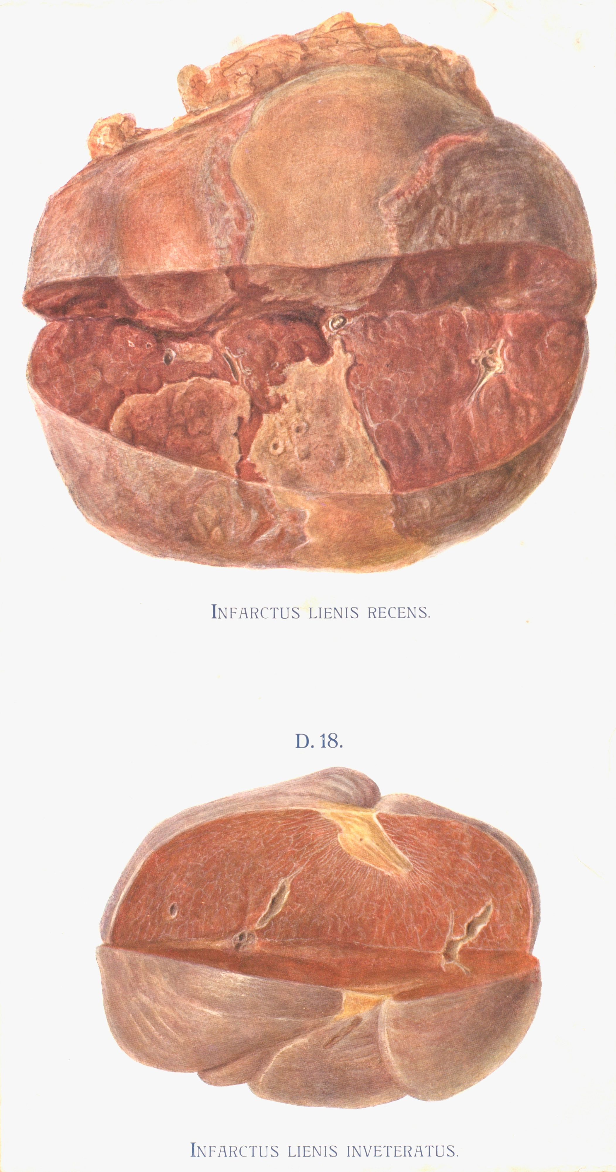
Plate #3 from Robert Knox’s “Man: His Structure & Physiology: Popularly Explained and Demonstrated”, 2nd edition, 1858.

Version 2

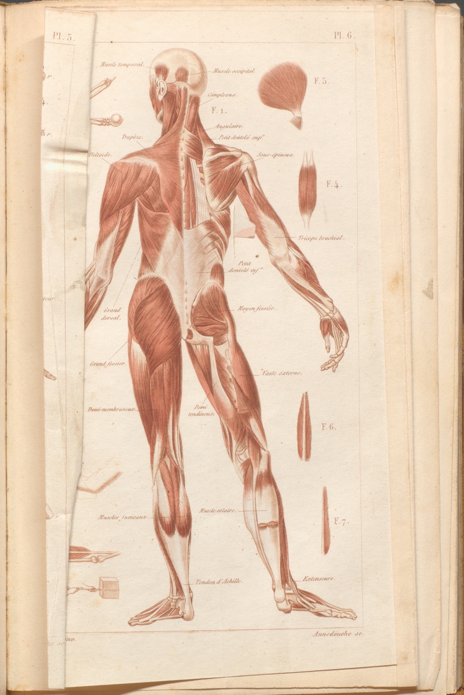
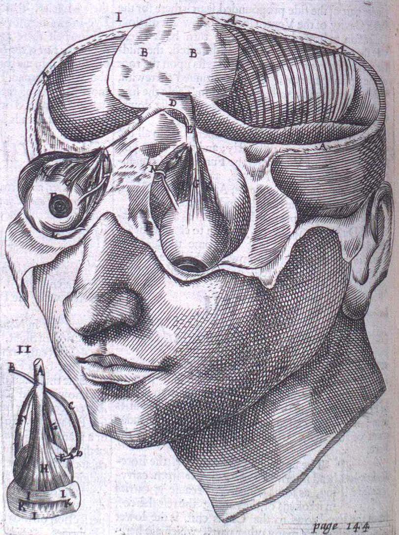
Plate #6 from Robert Knox’s “Man: His Structure & Physiology: Popularly Explained and Demonstrated”, 2nd edition, 1858.


pic_2016-07-19_171859 (1)

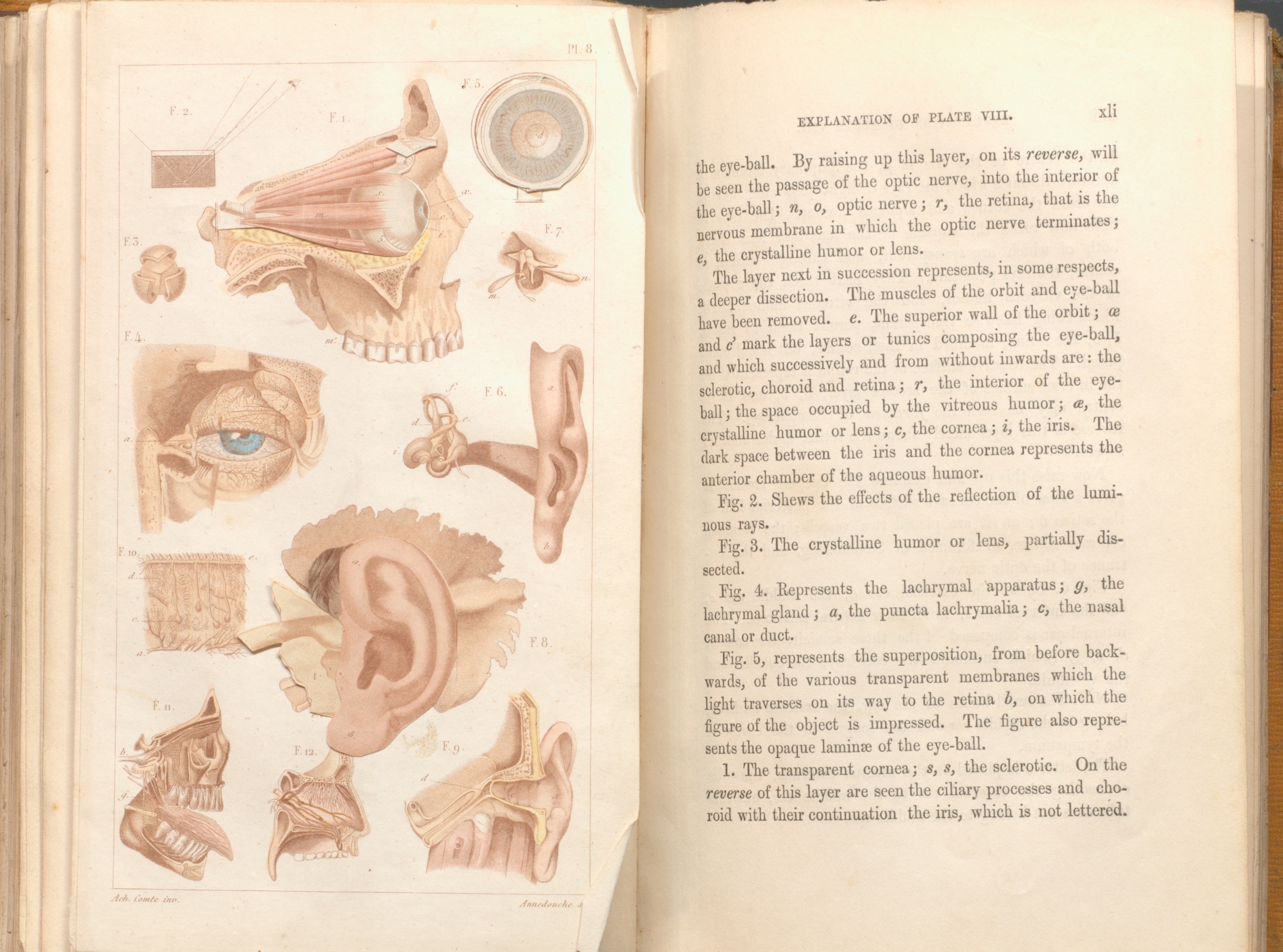
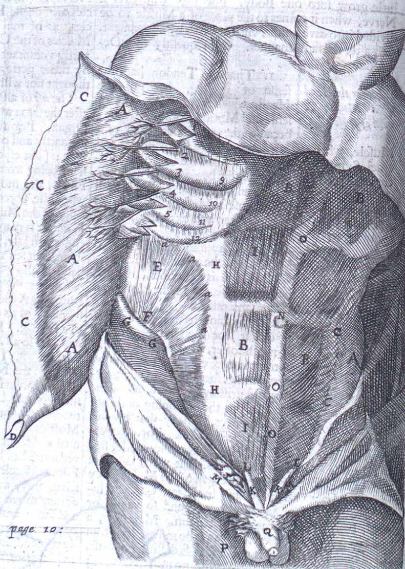
Plate #8 with explanations on opposite page in Robert Knox’s “Man: His Structure & Physiology: Popularly Explained and Demonstrated”, 2nd edition, 1858.

Pathologisch-anatomische Tafeln

pic_2016-06-13_151829

Illustration from Pathologisch-anatomische Tafeln: nach frischen Präparaten mit erläuterndem anatomisch-klinischem text, circa 1892

Recent summer housekeeping has turned up a series of rare mounted chromographed plate illustrations from late-19th century patho-anatomical atlas Pathologisch-anatomische Tafeln: nach frischen Präparaten mit erläuterndem anatomisch-klinischem text (Pathological anatomy plates: reproduced from fresh preparations with explanatory anatomical-clinical text) by German internist Alfred Kast (1856-1903).

The archives has eight of these illustrations in total, donated to the Osler Library back in the 1970s as part of the Maude Abbott Collection. Unfortunately no copy of this atlas exists at McGill, however WorldCat shows The Thomas Fisher Rare Book Library at the University of Toronto as having a copy in their catalogue. For those interested in seeing more of this series, The Wellcome Trust in London has fifteen of Kast’s anatomical illustrations included in their online Wellcome Images collection.

pic_2016-06-06_144022

Illustration with microscopic detail from Pathologisch-anatomische Tafeln: nach frischen Präparaten mit erläuterndem anatomisch-klinischem text, circa 1892

pic_2016-06-06_144056

Illustration with microscopic detail from Pathologisch-anatomische Tafeln: nach frischen Präparaten mit erläuterndem anatomisch-klinischem text, circa 1892


pic_2016-06-06_144154

Illustration from Pathologisch-anatomische Tafeln: nach frischen Präparaten mit erläuterndem anatomisch-klinischem text, circa 1892

pic_2016-06-13_151812

Illustration from Pathologisch-anatomische Tafeln: nach frischen Präparaten mit erläuterndem anatomisch-klinischem text, circa 1892

Preserving the “art” in Heart

pic_2016-06-15_134036

A sampling of Maude Abbott’s wooden printing blocks, housed at Osler Library.

Last week in the archives, we made a few updates to our Maude Abbott Collection that included rehousing and preserving various media and artifacts. For students who are unfamiliar, or are discovering the work of Dr. Maude Elizabeth Abbott (1869-1940) for the first time, we encourage you to explore the Osler Library’s collection.

Maude Abbott was a true medical luminary and a pioneer for women in medicine. Her extraordinary career garnered an international reputation for excellence in pathology, as well as medical museums – particularly impressive considering she was denied entry as a young medical student to McGill’s Faculty of Medicine in 1889 because of her gender (women were not admitted to the Faculty until 1917).

William Osler was one of Abbott’s greatest mentors, and was the first to encourage her research into congenital cardiac disease. These wooden printers blocks (above) and glass lantern slides (below) represent a small sampling of the many diagrams and illustrations created for Abbott’s ground-breaking Atlas of Congenital Cardiac Disease, published in 1936.

pic_2016-06-15_133453

A sampling of Maude Abbott’s glass slides, housed at Osler Library.

Bartholinus Anatomy

Born into a Danish family of physicians, Thomas Bartholin (1616-1680) was influenced at an early age by his relatives’ forays into anatomical science and medicine. He traveled widely throughout Europe before receiving his medical degree at Basel in 1645, and subsequently made significant contributions to 17th century anatomical knowledge, particularly concerning the discovery of the lymph vessel system as well as first recognizing the thoracic duct in humans. A prolific writer, his work was published both in Leiden and London and translated into several different languages. Like his father, Caspar Bartholin the Elder, Thomas wrote several anatomical treatises which became rather popular textbooks. This 1668 English translation of his most famous work, Bartholinus Anatomy, has recently made its way into the Osler Library. Published in London by Nicholas Culpeper and Abdiah Cole, the anatomical treatise contains 153 copperplate engravings (4 of which are fold-out plates).

pic_2013-07-26_100446_Part5

The text is a later adaptation of his father’s Anatomicae Institutiones Corporis Humani (1611), which was, for many years, a standard textbook on the subject of anatomy. Caspar’s work was revised and illustrated by Thomas who published the new edition, Anatomia, in 1641. Following his observations on the human lymphatic system (along with some criticism from fellow anatomists) Bartholin updated his Anatomia, publishing a revised and augmented edition in 1655.

The younger Bartholin’s textbook differs from that of his father with the addition of references to the writings of contemporary anatomists, such as William Harvey and Gasparo Aselli. The work of these two physicians is explicitly referenced in the book’s appendix: two letters from Johannes Walaeus to the author, “Concerning the Motion of the Chyle and Blood” and “Of the Motion of the Blood.” The accompanying illustrations likewise updated the original edition. These engraved plates, roughly inserted within and often overlapping the book’s text, supplement Bartholin’s discussion of the human body. Most of these images were not original, but rather were based on the work of Vesalius, Casserio, Vesling, and other famous anatomists.

pic_2013-07-29_160054_Part2

Bartholin’s Anatomy was one of the most popular anatomical texts of the 17th century. Separated into four books and four “petty books” detailing distinct sections and systems of the human body, the exhaustive work was central to the early modern study and development of the anatomical sciences.


Anatomical atlas donated in honour of outgoing principal

This Tuesday, April 30th, at Osler, Principal Monroe-Blum was presented with a significant rare work donated in her honour. The Exposition anatomique de la structure du corps human by Jacques Fabien Gautier d’Agoty (1716-1785) was published in France in 1759. D’Agoty was an artist who trained in colour printing with Jacob Christoph Le Blon (1670-1741), a German painter and engraver who developed the technique of colour mezzotint printing. D’Agoty took on the difficult and elaborate project of printing a complete, life-sized anatomy in colour. The resulting book is an elephant folio with nineteen pages of text and twenty colour mezzotint plates.

PrincipalsEvent PrincipalsEvent4

Mezzotint is an intaglio printing technique, meaning that a design is incised into a surface and the resulting image is created by the ink in the grooves. In mezzotint printing, the negative space in the image on the plate is roughened up and pitted with a tool called a rocker in order to achieve half-tones and shading. Le Blon’s colour mezzotint process involved making multiple engravings, one for each colour of ink, and then overlaying them. His original technique involved the use of red, blue, and yellow inks to create a range of colours and he later added a fourth layer of black.

dagoty PrincipalsEvent3

This copy of the Exposition anatomique now held at Osler is among only a handful of existing copies. Others are held in the Bibliothèque nationale de France, Yale University, Harvard University, and the University of Edinburgh, among others. It is also the only known copy in Canada. The atlas was acquired thanks to several generous donors and presented in recognition of Professor Heather Munroe-Blum’s ten years as Principal and Vice-Chancellor.

Photos: Sabrina Hanna