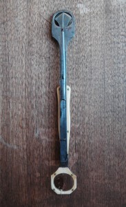
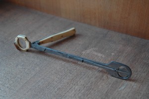
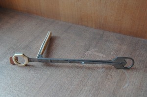
This tonsil guillotine (otherwise known as a tonsillotome) is one of hundreds of relics of the history of medicine housed in the Osler Library artifact collection.
Developed in the decades following the French Revolution, the tonsil guillotine has one glaring similarity to its much more sinister cousin: a sharp, fast-moving blade designed to cut the afflicted mass from its bulk. This much smaller blade is fastened in between two fixed steel plates, and attached to a moveable handle that slides it through an auxiliary ring intended to fit around the infected tonsil.
The instrument was originally developed as an adaptation to Benjamin Bell’s (1749-1806) uvulotome, which was similarly used to excise an inflamed and elongated uvula. Bell describes the use of this tool in his System of Surgery, “that part of the uvula intended to be removed being passed thro the opening of the body of the instrument, the cutting slider, which ought to be very sharp, must be pressed forward with sufficient firmness for dividing it from the parts above.”
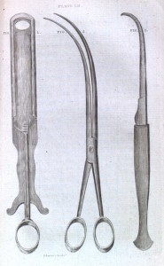
Instruments for removing the uvula and small tumors from the throat, from Benjamin Bell’s System of Surgery. 3rd ed. Edinburgh: C. Elliot & T. Kay, 1789.
The physician Philip Syng Physick (1768-1837) used this same device in treating a patient with a relentless cough in the spring of 1826. After noting the patient’s elongated uvula, Physick followed a popular treatment that involved partly removing the infected area. Up until that point, he, among other physicians, had used scissors or ligatures for such operations. Dr. Physick instead decided to try an old instrument in order to make the process easier. His design was very similar to that of Bell’s uvulotome: a sharp sliding blade would pass through two plates into a round opening at the end of the instrument to remove the uvula in one smooth motion. In subsequent years Physick adapted this instrument for use in excising infected tonsils, enlarging the aperture of the ring and making slight modifications to the steel body. The guillotine in our artifact collection follows Physick’s early design of a pointed and movable cutting blade between two steel plates. On one end of the device is a bone handle and ring used to position and slide the blade within the patient’s mouth. The steel hoop at the opposite end of the instrument may have been covered by a strip of wax linen to achieve a cleaner cut, and the thin needle lying flat above the blade would have kept the infected tonsil in place during the operation.
 At this time, tonsillectomies were considerably restricted by inadequate anesthetic, so surgeons made every effort to perform the operation as quickly as possible. This is perhaps why the guillotine became a popular tool for the operation; contemporary alternatives involved the use of curved scissors (which often led to excessive hemorrhaging) or the use of a wire ligature to slowly separate the tonsil from the inside of the mouth (a long and excruciating process without viable painkillers). In contrast, the guillotine could perform the excision in one even movement.
Further modifications of the tonsil guillotine were made by Morel Mackenzie in the 1860s, popularizing the instrument for wider use. It remained the preferred method for tonsillectomy until the early twentieth century, until it became more common to perform complete rather than partial removal of the tonsil. A technique involving removal of the tonsil with a scalpel and forceps proved much more effective and precise, and tonsillectomy using the guillotine eventually fell out of favor with most physicians.
References
Benjamin Bell. A System of Surgery. Edinburgh: C. Elliot & T. Kay, 1789.
J. Mathews, J. Lancaster, I. Sherman, and G. O. Sullivan. “Guillotine tonsillectomy: a glimpse into its history and current status in the United Kingdom.” The Journal of Laryngology & Otology 116 (Dec. 2002): 988–991.
Neil G. McGuire. “A method of guillotine tonsillectomy with an historical review.” The Journal of Laryngology & Otology 81, no. 2 (Feb. 1967): 187-195.
Ronald Alastair McNeill. “A History of Tonsillectomy: Two Millenia of Trauma, Haemorrhage and Controversy.” The Ulster Medical Journal 29, no. 1 (June 1960): 59-63.
Philip Syng Physick. “Case of Obstinate Cough, occasioned by elongation of the Uvula, in which a portion of that organ was cut off, with a description of the instrument employed for that purpose, and also for excision of scirrhous tonsils.” The American Journal of the Medical Sciences 1, no. 2 (1828): 262-265.


