Découvrir l’anatomie grâce aux livres animés

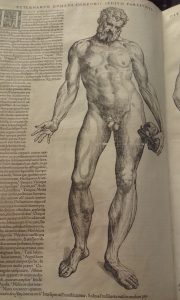
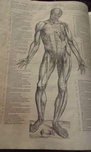
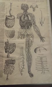
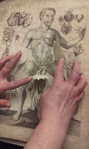
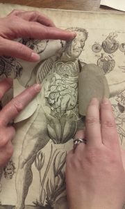
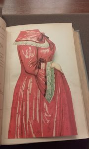
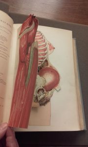
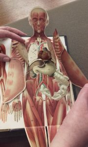
Pratiquée de manière assez confidentielle au Moyen Age, la dissection se développe au 16e siècle, notamment lorsque le pape Clément VII autorise son enseignement. La Renaissance est également marquée par un essor des livres dédiés à l’anatomie, et en particulier des livres animés. Les auteurs de l’époque rivalisent d’imagination et d’ingéniosité pour répondre à la soif de connaissance des curieux, des médecins et des étudiants. Ces ouvrages ont souvent un objectif pédagogique, ce qui explique le recours aux illustrations à volets qui permettent au lecteur de mieux visualiser l’organisation interne du corps humain: grâce à un système de volets, le lecteur peut en quelque sorte pratiquer lui-même une dissection en soulevant un à un les volets qui dévoilent à chaque fois une « couche » du corps humain.

Parmi les anatomistes du 16e siècle, le médecin bruxellois André Vésale est sans doute le plus connu. Convaincu qu’il est difficile d’enseigner et d’apprendre l’anatomie sans pouvoir observer directement le corps humain, Vésale pratique régulièrement la dissection et fait paraître en 1543  son De humani corporis fabrica, qui renouvelle les connaissances en anatomie depuis Galien.

Dans son Epitome, supplément à la Fabrica destiné essentiellement aux étudiants en médecine, Vésale propose lui aussi une sorte de livre animé : plusieurs planches anatomiques de l’Epitome peuvent être découpées par le lecteur afin de reconstituer in fine un corps humain en trois dimensions. En effet, comme l’explique Jacqueline Vons, l’Epitome contient « deux planches constituées de plusieurs dessins que l’étudiant doit découper et coller sur d’autres figures afin d’obtenir, par superposition, un corps humain en trois dimensions. Le texte accompagnant ces deux planches est l’équivalent d’un mode d’emploi, d’une notice qui doit guider pas à pas l’étudiant dans son travail de recomposition. C’est déjà une méthode pédagogique inter-active, qui consiste à s’adresser à un étudiant virtuel, en développant une forme de dialogue entre le maître et le disciple » (1).

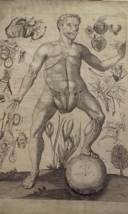
Un autre exemple célèbre de livre animé est le Pinax microcosmographicus de Johann Remmelin, paru au début du 17e siècle et réédité à plusieurs reprises (la bibliothèque Osler en possède d’ailleurs deux exemplaires du début du 18e siècle). Il permet de découvrir l’anatomie masculine et féminine grâce à des illustrations à volets. La bibliothèque de l’université de Columbia a récemment numérisé cet ouvrage et a mis en ligne une vidéo très intéressante qui montre le fonctionnement de ces illustrations anatomiques à volets.

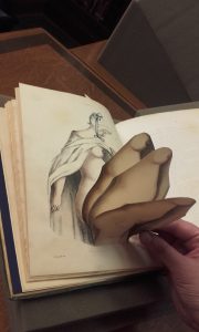
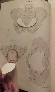
La pratique du livre anatomique à volets se perpétue et atteint son apogée au 19e siècle, quand les techniques d’impression en couleur se perfectionnent et se développent. Nous vous présentons ici quelques exemples issus de nos collections.

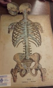
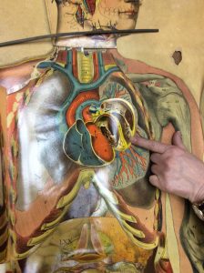
Mais les illustrations à volets ne se limitent pas au livre : certaines planches anatomiques utilisent le même procédé. La bibliothèque Osler a par exemple fait récemment l’acquisition d’une planche anatomique sur bois du 19e siècle, sans doute utilisée pour l’enseignement, qui représente un corps masculin presque à taille réelle, et qui par un système de volets maintenus par des languettes amovibles en métal, permet d’avoir la vision interne du corps humain (muscles, tendons, organes, os…).

Notes:

(1) Vons J., « L’Epitome, un ouvrage méconnu d’André Vésale (1543) », Histoire des sciences médicales, XL, 2, 2006, p.177-189, disponible en ligne [consulté le 12/04/2017].


Bibliographie :

Binet J.-L., « Traités d’anatomie », Bibliothèque numérique Medic@, disponible en ligne [consulté le 19/04/2017].

Carlino A., Paper bodies: a catalogue of anatomical fugitive sheets (1538-1687), London, Wellcome Institute for the History of Medicine, 1999.

Columbia University Medical Center, « A medical pop-up from the 17th century”, disponible en ligne [consulté le 19/04/2017].

Duke University Libraries, « Animated anatomy », 2011, disponible en ligne [consulté le 12/04/2017].

Van Wijland J., « Usages des papiers découpés dans l’illustration anatomique des XVIe-XVIIe siècles », disponible en ligne [consulté le 19/04/2017].

Vons J., « L’Epitome, un ouvrage méconnu d’André Vésale (1543) », Histoire des sciences médicales, XL, 2, 2006, p.177-189, disponible en ligne [consulté le 12/04/2017].

Vons J., « L’anatomie au 16e siècle », Bibliothèque numérique Medic@, disponible en ligne [consulté le 19/04/2017].


Dissection Room Records 1883-1908

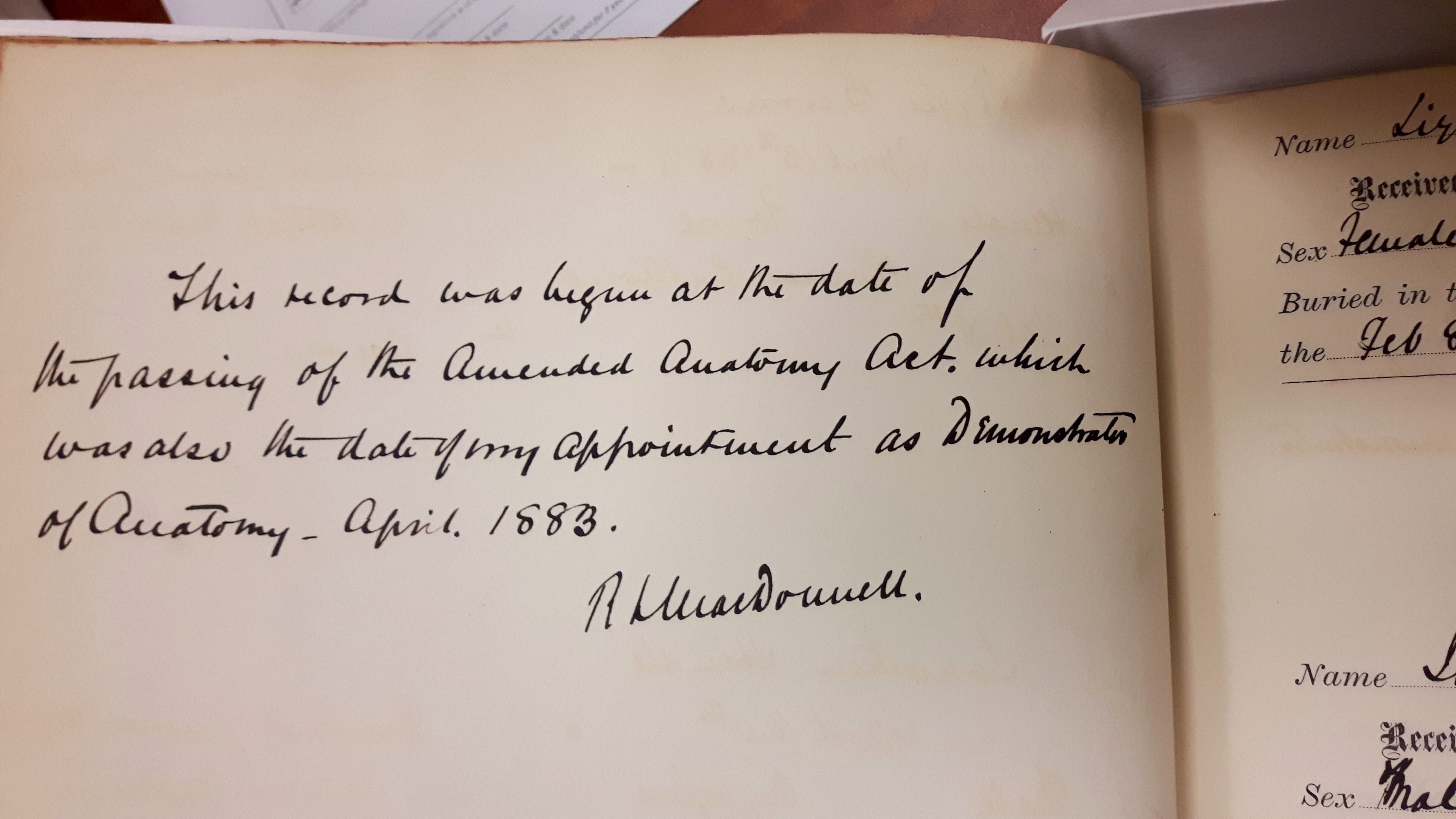
Inscription on first page of Dissection Room Record 1883-1891 written by Dr. Richard Lea MacDonnell, Demonstrator of Anatomy, McGill University in April 1883.

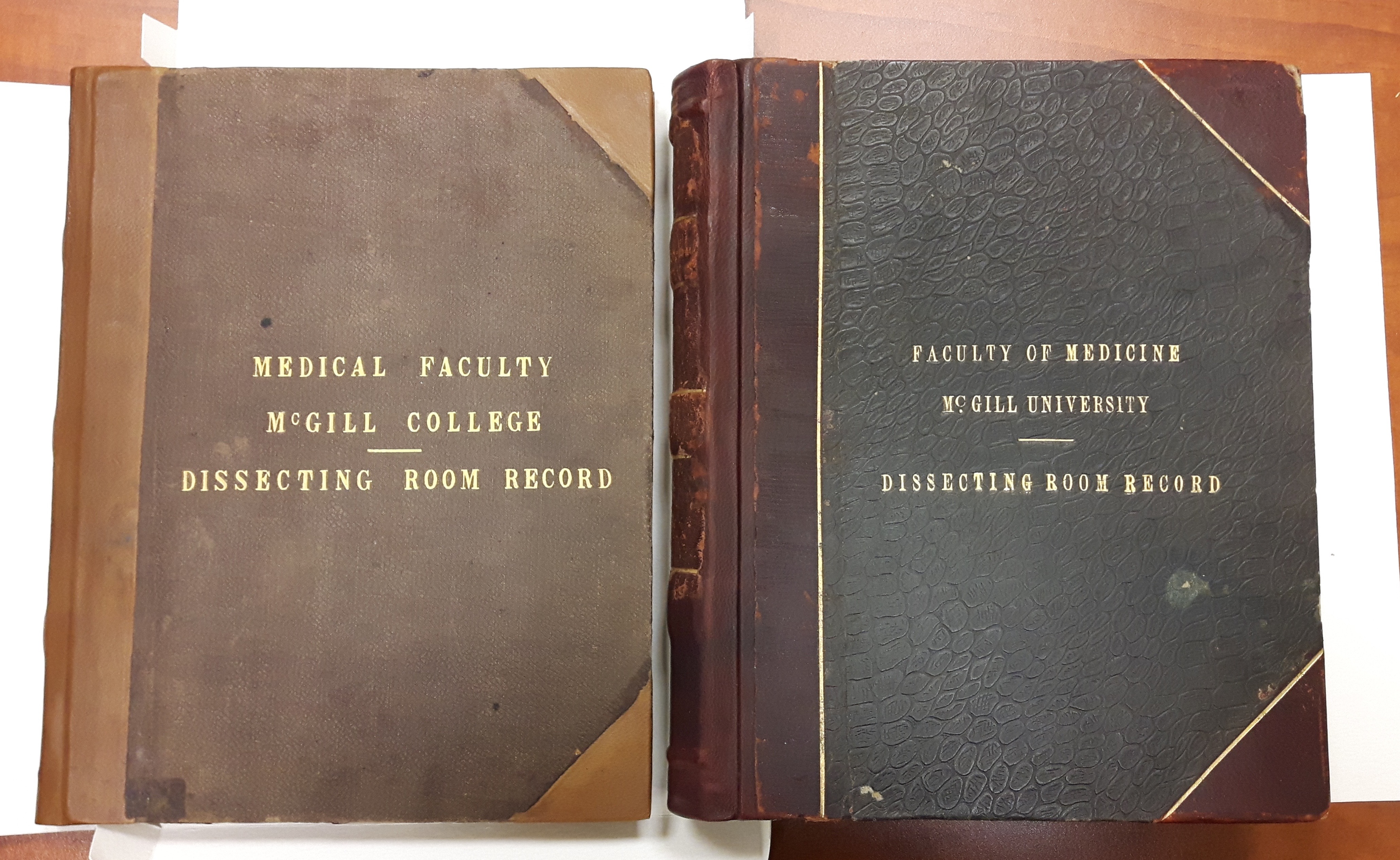
We are pleased to have these historical records back at the Osler Library after receiving recent conservation treatment. These books contain records of all McGill Faculty of Medicine dissection cadavers in the Department of Anatomy from 1883-1891, and 1896-1908.

When the Legislative Assembly of the Province of Canada’s Anatomy Act of 1843 was amended in April 1883, Senior Demonstrator of Anatomy Dr. Richard Lea MacDonnell began keeping these detailed records at McGill. Prior to the Anatomy Act, body-snatching was a nefarious problem often associated with the study of anatomy. The 1843 and 1883 Acts allowed for more corpses to be made available to medical schools for the purpose of teaching and learning. The records preserved within these books provide details and evidence of the Department of Anatomy’s legally acquired cadavers at the time. Each entry includes details such as name, sex, age, cause of death, religion, date received, which hospital the cadaver was received from, and the date and location of burial.

Richard Lea MacDonnell (1856-1891) was the son of Dublin surgeon Dr. Robert Lea MacDonnell. A graduate of McGill in 1876, McDonnell went on to become a prominent figure within the Faculty of Medicine before his untimely death at the age of 35. William Osler paid tribute to his friend and colleague in the New York Medical Journal, writing: “Although only thirty-five years old, he [MacDonnell] had reached a position which gave scope to abilities of first-class order and afforded opportunities of impressing upon a large class of students those qualities of mind so essential in the teacher, so priceless to the taught – honesty, system, and painstaking care” (NYMJ, 54: 162, 1891).

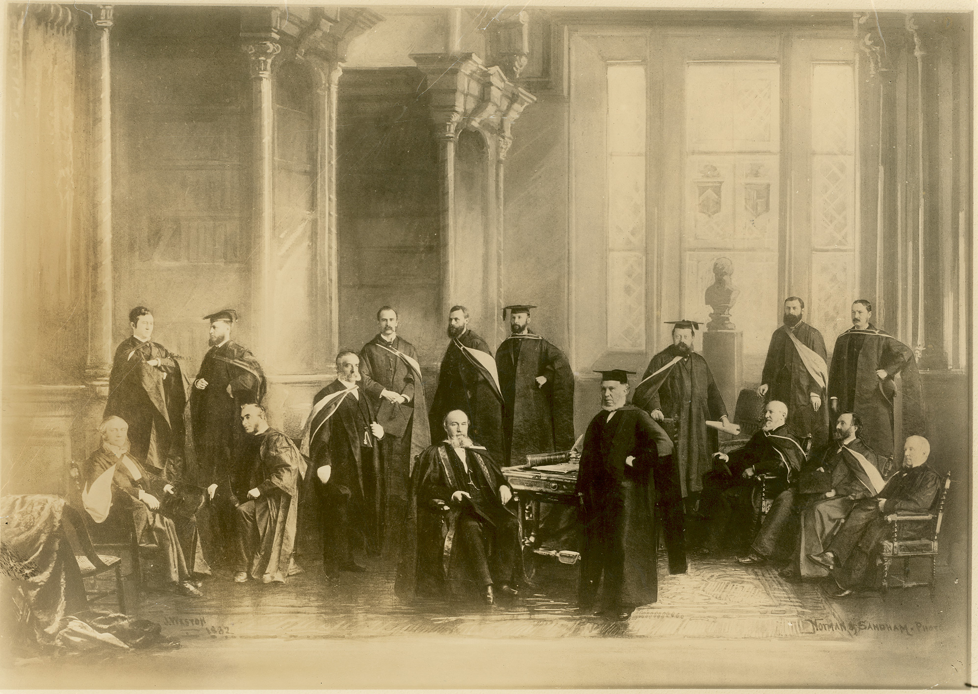
Below is a composite portrait of McGill Faculty of Medicine in 1882 from our William Osler Photo Collection. William Osler is standing fourth from left, and Richard Lea MacDonnell stands on the far right. A new Richard L. MacDonnell Collection (P133) has been created in the Osler Library Archives, and these dissection books along with several fascinating scrapbooks put together by MacDonnell are now available to view upon request.

“McGill University Faculty of Medicine at its Semicentennial, 1882”, William Osler Photo Collection, Osler Library of the History of Medicine, CUS_033-011_P. Standing, from left to right, are Thomas G. Roddick, George Ross, William E. Scott, William Osler, Francis J. Shepherd, William Gardner, George W. Campbell, Gilbert Prout Girdwood, Frank Buller, and Richard L. MacDonell. Sitting, from left to right, are Robert Palmer Howard, William Wright, John William Dawson, Duncan C. MacCallum, Robert Craik, and George E. Fenwick.

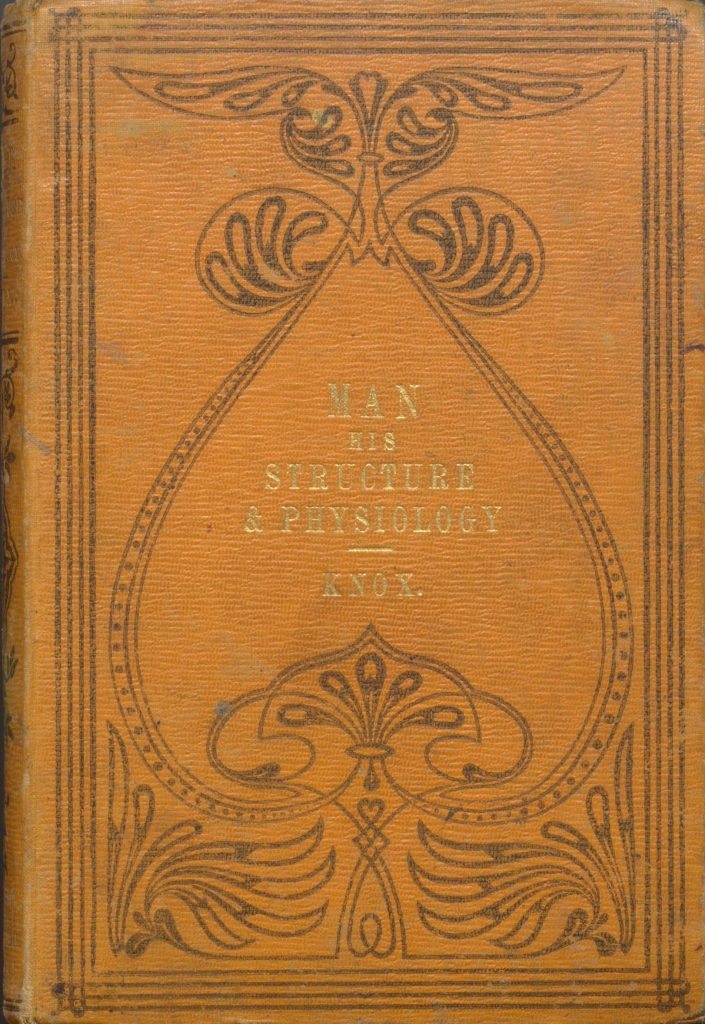
Man: His Structure & Physiology

pic_2016-07-19_171814

“Man: His Structure & Physiology: Popularly Explained and Demonstrated” by Robert Knox, 2nd edition, London: H. Bailliere, 1858.

This month we’ve chosen to highlight an anatomical atlas by Scottish anatomist, zoologist, and physician, Dr. Robert Knox (1791-1862). His popular book entitled Man: His Structure & Physiology: Popularly Explained and Demonstrated was originally published in 1857, with a second edition (shown here) printed a year later in 1858.

Knox was an esteemed professor at The University of Edinburgh — famous for his dissections and lectures which were often ticketed and open to the public. Prior to the 1832 Anatomy Act, it was discovered that Knox relied on illegal methods to acquire his cadavers. Knox was connected to the Burke and Hare West Port murders of 1828, and despite never being tried, his reputation was forever marred in controversy.

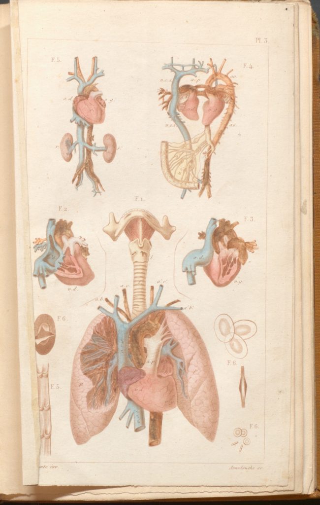
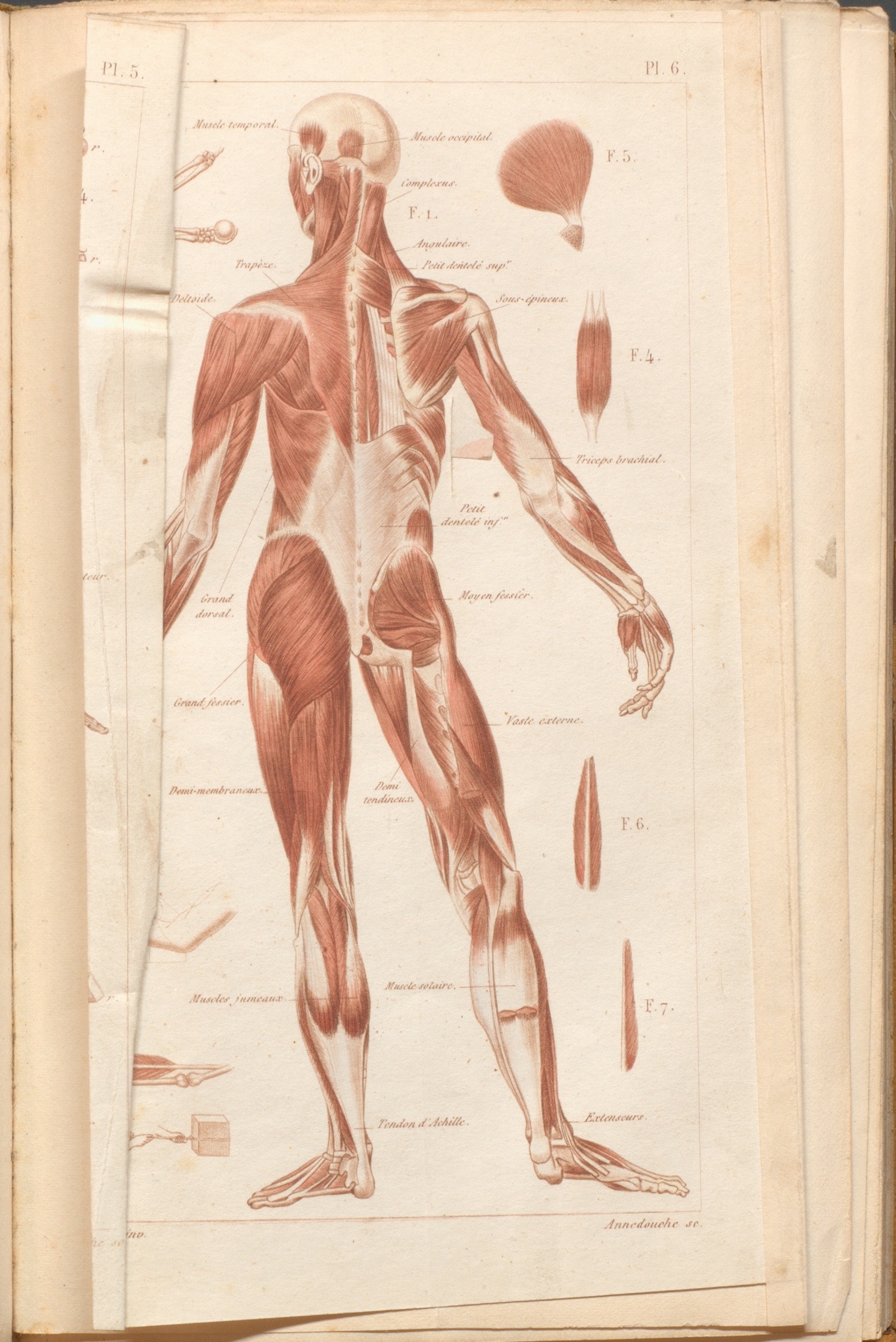
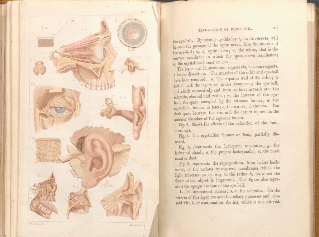
The atlas is described in simple language and includes some detailed plate illustrations — several of which can lift (“pop-up”) off the page. The idea behind this design was to imitate a dissection as much as possible, allowing students and readers to discover multiple layers of physiological detail. As the preface of the second edition describes, it is an “elementary and educational Work, containing such an outline of Human Structure and Human Physiology as may prove a safe basis whereon to build the edifice of special or philosophic inquiry and research” (London, October, 1857).

The book is available to view at the Osler Library during regular hours. For those who are not able to visit the library in person, a digitized version of a more recent pressing can be accessed at archive.org.

Version 2

Plate #3 from Robert Knox’s “Man: His Structure & Physiology: Popularly Explained and Demonstrated”, 2nd edition, 1858.

Version 2

Plate #6 from Robert Knox’s “Man: His Structure & Physiology: Popularly Explained and Demonstrated”, 2nd edition, 1858.


pic_2016-07-19_171859 (1)

Plate #8 with explanations on opposite page in Robert Knox’s “Man: His Structure & Physiology: Popularly Explained and Demonstrated”, 2nd edition, 1858.

Pathologisch-anatomische Tafeln

pic_2016-06-13_151829

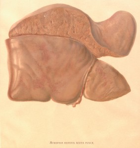
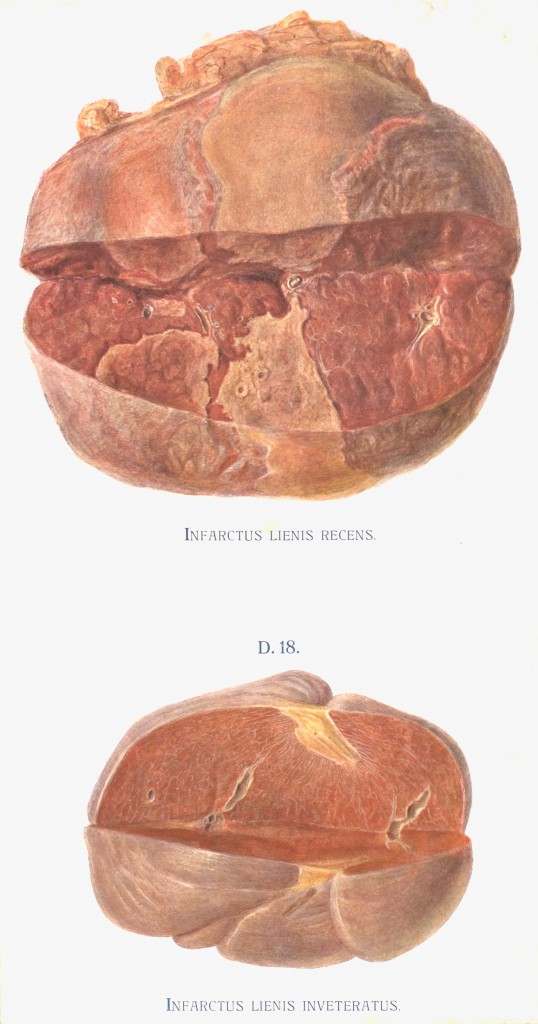
Illustration from Pathologisch-anatomische Tafeln: nach frischen Präparaten mit erläuterndem anatomisch-klinischem text, circa 1892

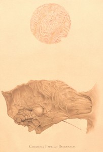
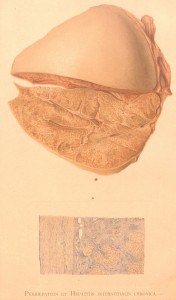
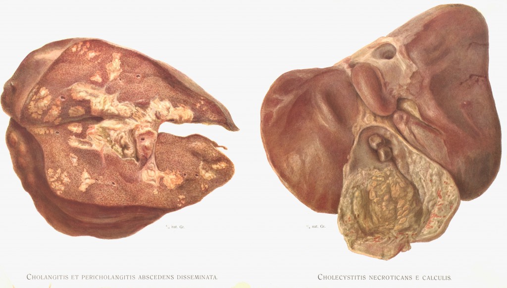
Recent summer housekeeping has turned up a series of rare mounted chromographed plate illustrations from late-19th century patho-anatomical atlas Pathologisch-anatomische Tafeln: nach frischen Präparaten mit erläuterndem anatomisch-klinischem text (Pathological anatomy plates: reproduced from fresh preparations with explanatory anatomical-clinical text) by German internist Alfred Kast (1856-1903).

The archives has eight of these illustrations in total, donated to the Osler Library back in the 1970s as part of the Maude Abbott Collection. Unfortunately no copy of this atlas exists at McGill, however WorldCat shows The Thomas Fisher Rare Book Library at the University of Toronto as having a copy in their catalogue. For those interested in seeing more of this series, The Wellcome Trust in London has fifteen of Kast’s anatomical illustrations included in their online Wellcome Images collection.

pic_2016-06-06_144022

Illustration with microscopic detail from Pathologisch-anatomische Tafeln: nach frischen Präparaten mit erläuterndem anatomisch-klinischem text, circa 1892

pic_2016-06-06_144056

Illustration with microscopic detail from Pathologisch-anatomische Tafeln: nach frischen Präparaten mit erläuterndem anatomisch-klinischem text, circa 1892


pic_2016-06-06_144154

Illustration from Pathologisch-anatomische Tafeln: nach frischen Präparaten mit erläuterndem anatomisch-klinischem text, circa 1892

pic_2016-06-13_151812

Illustration from Pathologisch-anatomische Tafeln: nach frischen Präparaten mit erläuterndem anatomisch-klinischem text, circa 1892

Anatomical atlas donated in honour of outgoing principal

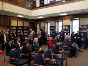
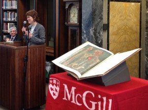
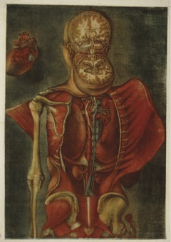
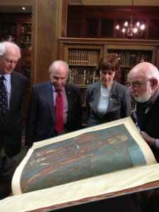
This Tuesday, April 30th, at Osler, Principal Monroe-Blum was presented with a significant rare work donated in her honour. The Exposition anatomique de la structure du corps human by Jacques Fabien Gautier d’Agoty (1716-1785) was published in France in 1759. D’Agoty was an artist who trained in colour printing with Jacob Christoph Le Blon (1670-1741), a German painter and engraver who developed the technique of colour mezzotint printing. D’Agoty took on the difficult and elaborate project of printing a complete, life-sized anatomy in colour. The resulting book is an elephant folio with nineteen pages of text and twenty colour mezzotint plates.

PrincipalsEvent PrincipalsEvent4

Mezzotint is an intaglio printing technique, meaning that a design is incised into a surface and the resulting image is created by the ink in the grooves. In mezzotint printing, the negative space in the image on the plate is roughened up and pitted with a tool called a rocker in order to achieve half-tones and shading. Le Blon’s colour mezzotint process involved making multiple engravings, one for each colour of ink, and then overlaying them. His original technique involved the use of red, blue, and yellow inks to create a range of colours and he later added a fourth layer of black.

dagoty PrincipalsEvent3

This copy of the Exposition anatomique now held at Osler is among only a handful of existing copies. Others are held in the Bibliothèque nationale de France, Yale University, Harvard University, and the University of Edinburgh, among others. It is also the only known copy in Canada. The atlas was acquired thanks to several generous donors and presented in recognition of Professor Heather Munroe-Blum’s ten years as Principal and Vice-Chancellor.

Photos: Sabrina Hanna


Exhibition catalogue now online

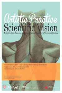
APposter1

The exhibition catalogue for our current exhibition, “Artistic Practice Scientific Vision: British Artistic Anatomy in the Late Eighteenth and Early Nineetenth Century,” curated by Dr. Allister Neher is now online!

This vivid exhibition explores the intersection of art and anatomy in late eighteenth and early nineteenth century Britain. Medical students and art students of the period frequented the same milieux, as aspiring doctors studied drawing at places like London’s Royal Academy of Arts and young artists studied anatomy at private medical schools.

Be sure not to miss the exhibition in person, on through the end of February.