Découvrir l’anatomie grâce aux livres animés

Pratiquée de manière assez confidentielle au Moyen Age, la dissection se développe au 16e siècle, notamment lorsque le pape Clément VII autorise son enseignement. La Renaissance est également marquée par un essor des livres dédiés à l’anatomie, et en particulier des livres animés. Les auteurs de l’époque rivalisent d’imagination et d’ingéniosité pour répondre à la soif de connaissance des curieux, des médecins et des étudiants. Ces ouvrages ont souvent un objectif pédagogique, ce qui explique le recours aux illustrations à volets qui permettent au lecteur de mieux visualiser l’organisation interne du corps humain: grâce à un système de volets, le lecteur peut en quelque sorte pratiquer lui-même une dissection en soulevant un à un les volets qui dévoilent à chaque fois une « couche » du corps humain.

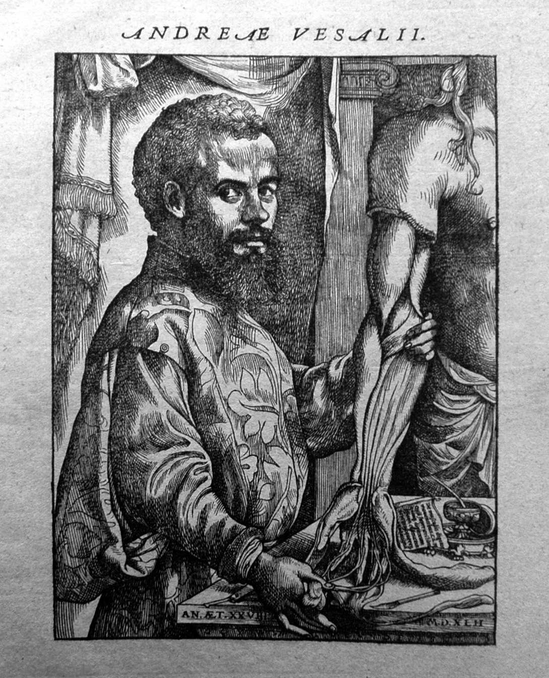
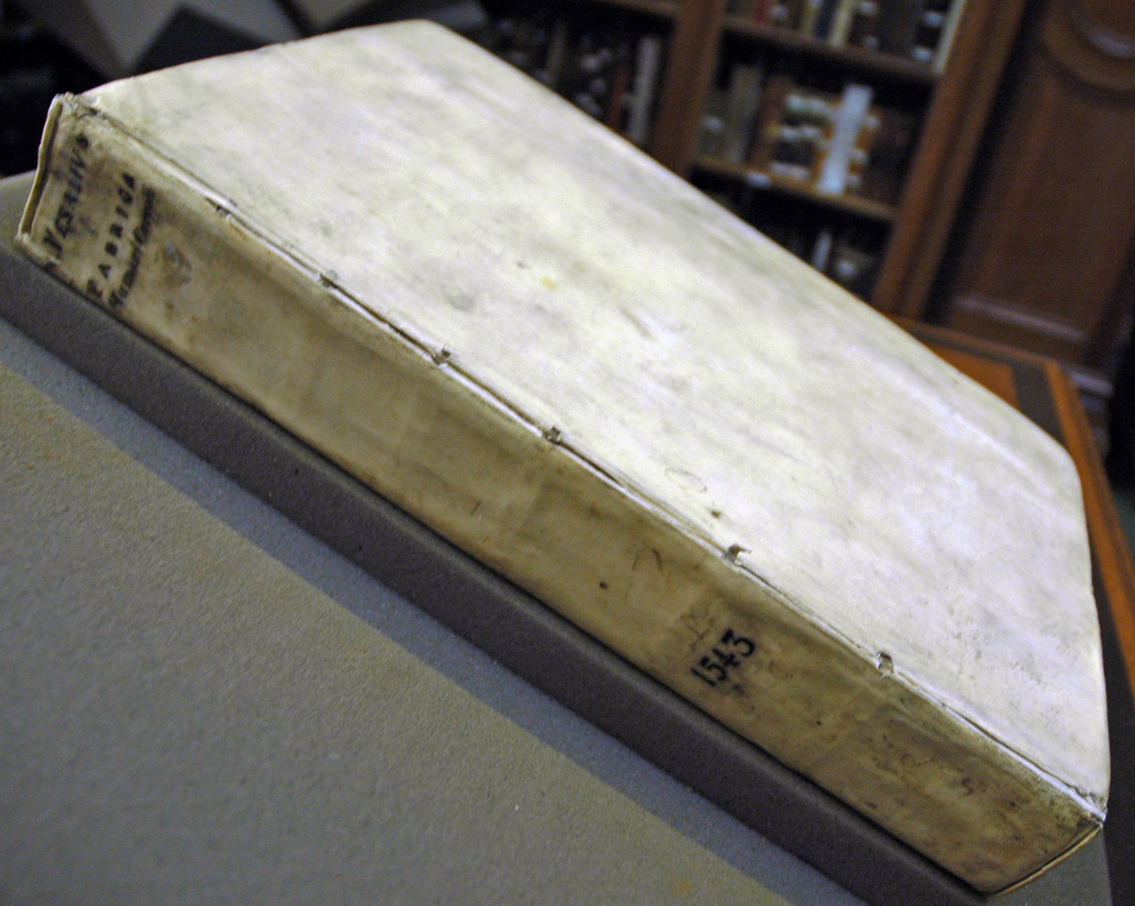
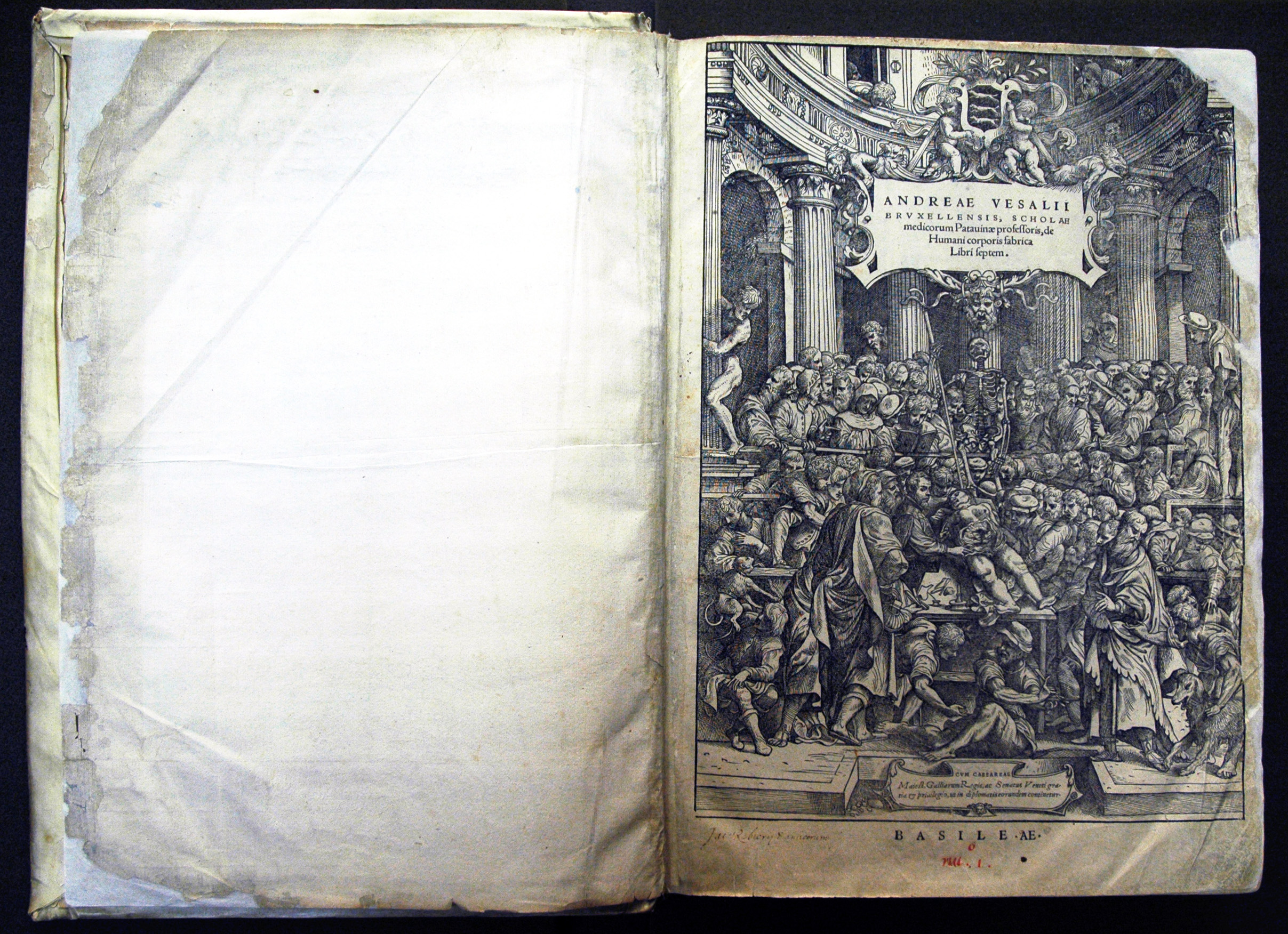
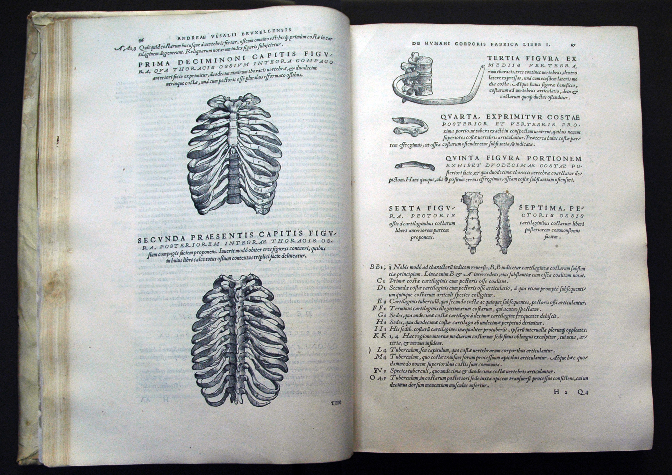
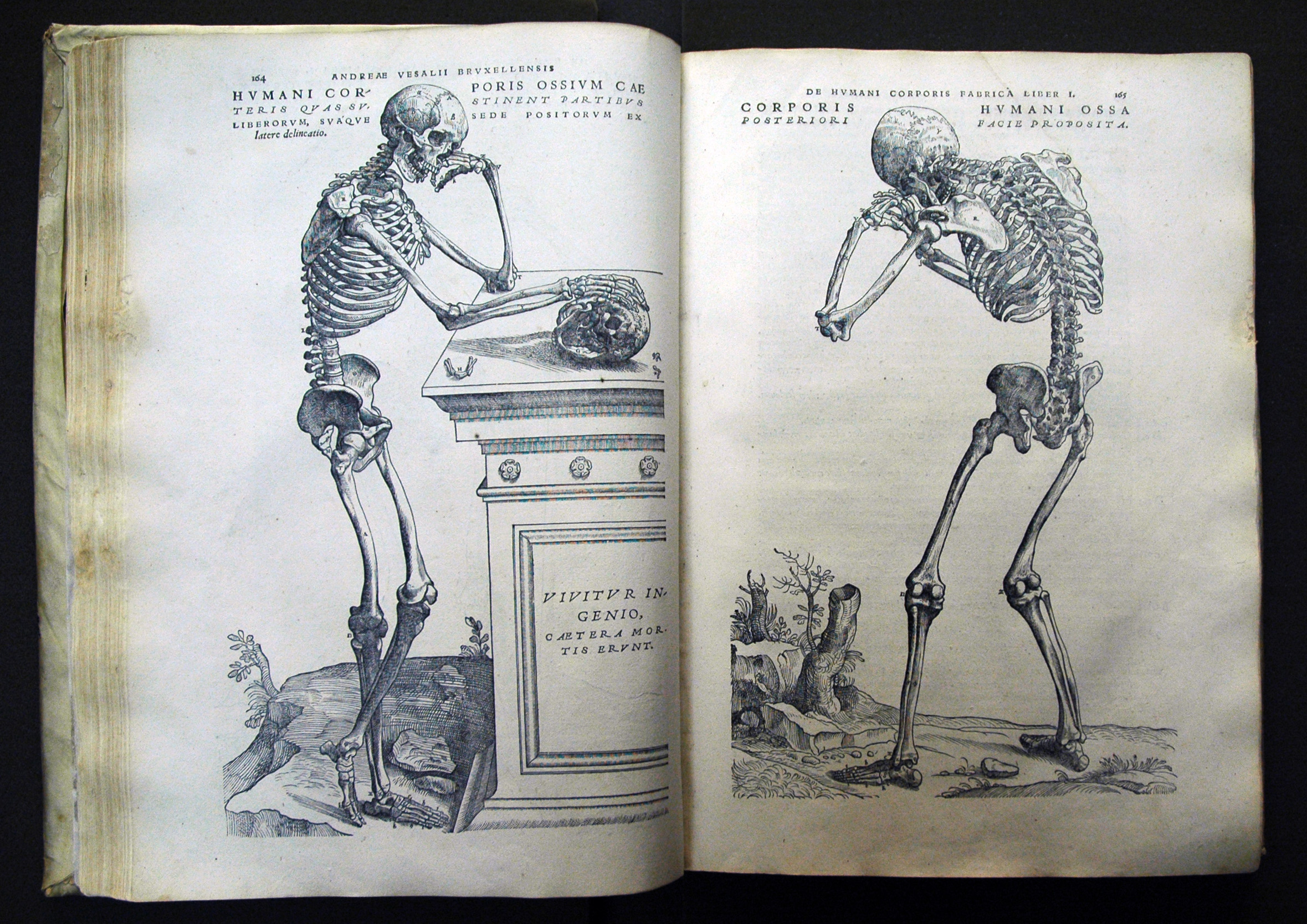
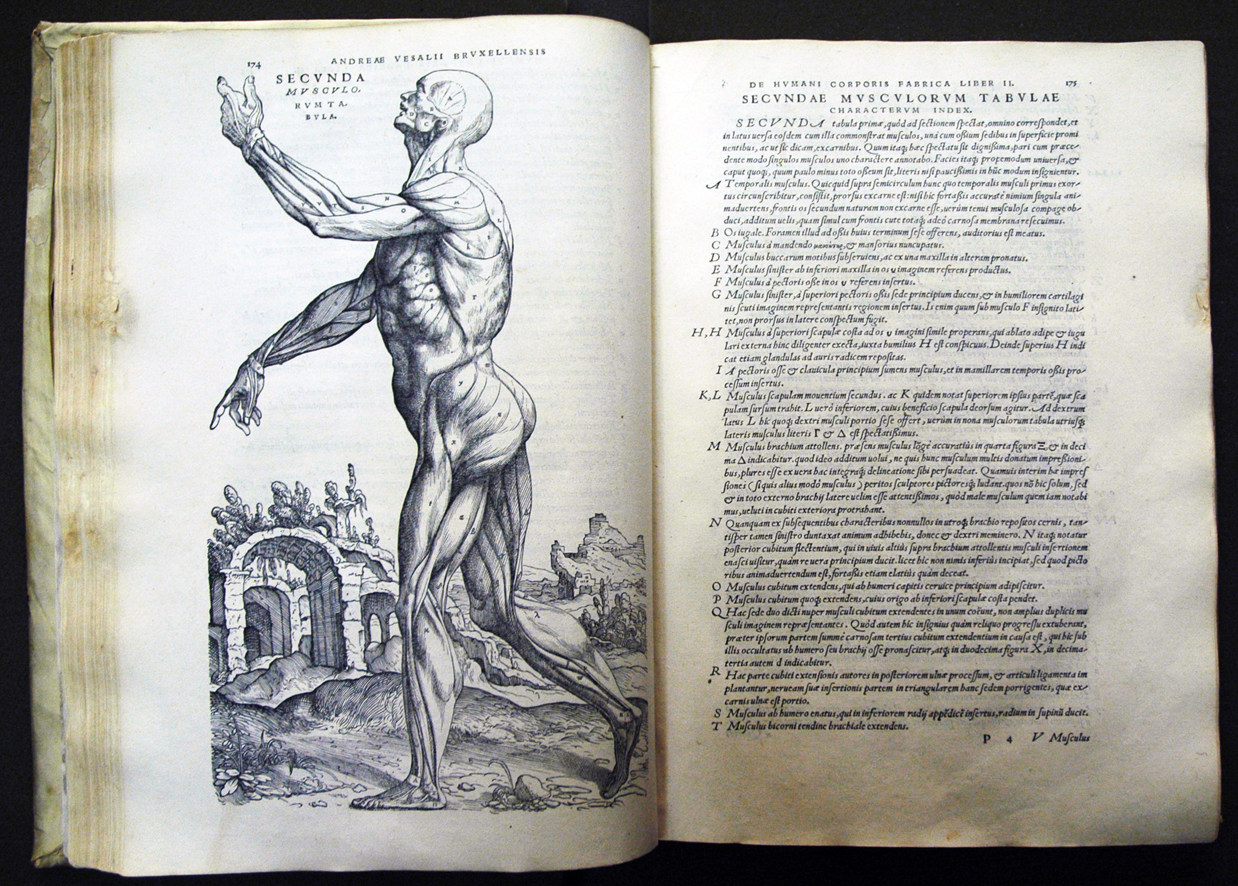
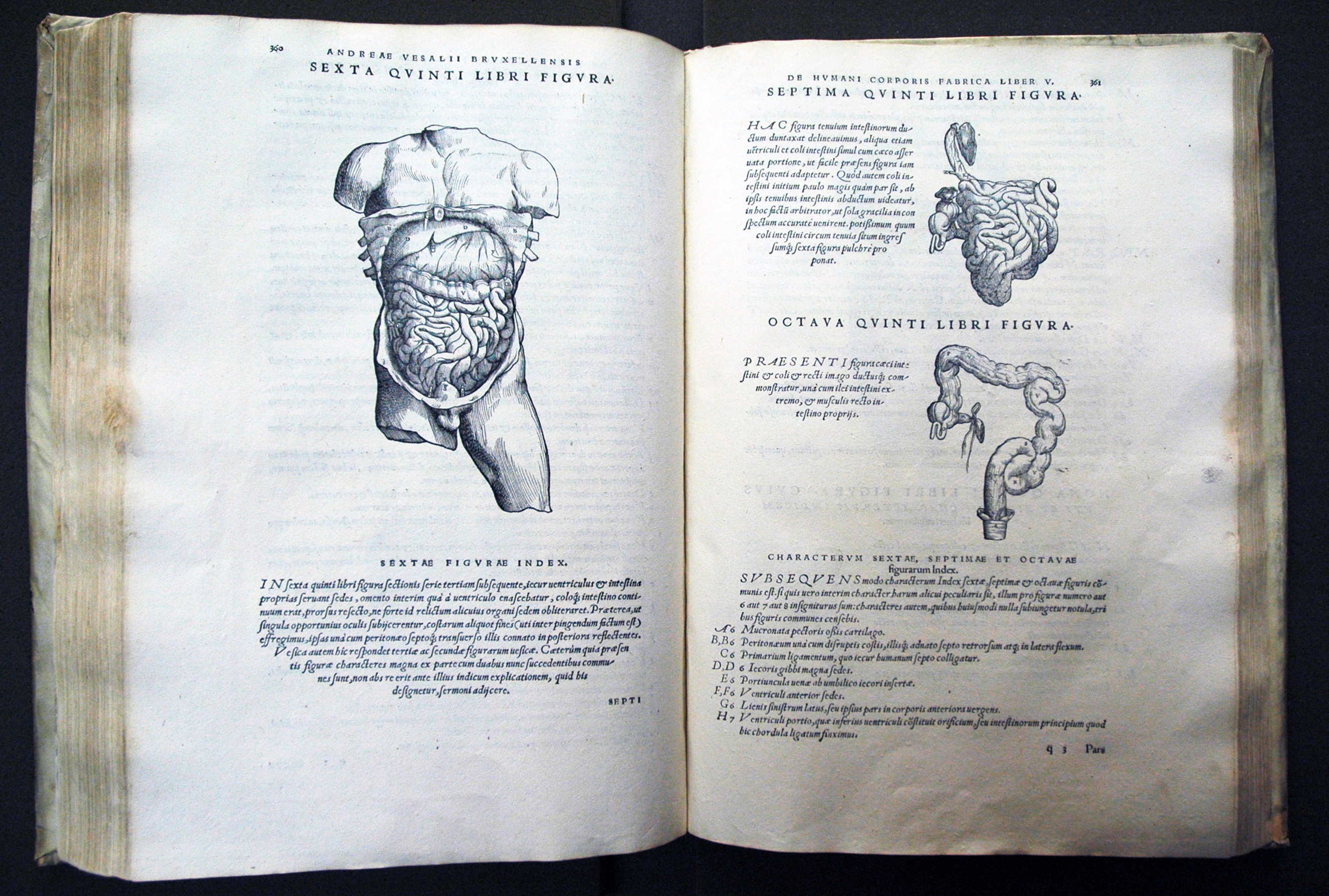
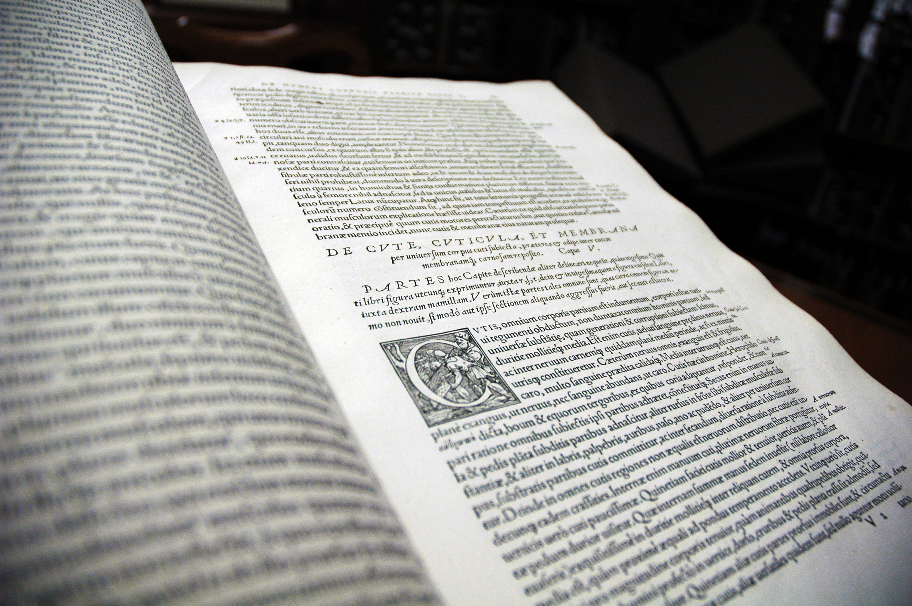
Parmi les anatomistes du 16e siècle, le médecin bruxellois André Vésale est sans doute le plus connu. Convaincu qu’il est difficile d’enseigner et d’apprendre l’anatomie sans pouvoir observer directement le corps humain, Vésale pratique régulièrement la dissection et fait paraître en 1543  son De humani corporis fabrica, qui renouvelle les connaissances en anatomie depuis Galien.

Dans son Epitome, supplément à la Fabrica destiné essentiellement aux étudiants en médecine, Vésale propose lui aussi une sorte de livre animé : plusieurs planches anatomiques de l’Epitome peuvent être découpées par le lecteur afin de reconstituer in fine un corps humain en trois dimensions. En effet, comme l’explique Jacqueline Vons, l’Epitome contient « deux planches constituées de plusieurs dessins que l’étudiant doit découper et coller sur d’autres figures afin d’obtenir, par superposition, un corps humain en trois dimensions. Le texte accompagnant ces deux planches est l’équivalent d’un mode d’emploi, d’une notice qui doit guider pas à pas l’étudiant dans son travail de recomposition. C’est déjà une méthode pédagogique inter-active, qui consiste à s’adresser à un étudiant virtuel, en développant une forme de dialogue entre le maître et le disciple » (1).

Un autre exemple célèbre de livre animé est le Pinax microcosmographicus de Johann Remmelin, paru au début du 17e siècle et réédité à plusieurs reprises (la bibliothèque Osler en possède d’ailleurs deux exemplaires du début du 18e siècle). Il permet de découvrir l’anatomie masculine et féminine grâce à des illustrations à volets. La bibliothèque de l’université de Columbia a récemment numérisé cet ouvrage et a mis en ligne une vidéo très intéressante qui montre le fonctionnement de ces illustrations anatomiques à volets.

La pratique du livre anatomique à volets se perpétue et atteint son apogée au 19e siècle, quand les techniques d’impression en couleur se perfectionnent et se développent. Nous vous présentons ici quelques exemples issus de nos collections.

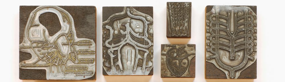
Mais les illustrations à volets ne se limitent pas au livre : certaines planches anatomiques utilisent le même procédé. La bibliothèque Osler a par exemple fait récemment l’acquisition d’une planche anatomique sur bois du 19e siècle, sans doute utilisée pour l’enseignement, qui représente un corps masculin presque à taille réelle, et qui par un système de volets maintenus par des languettes amovibles en métal, permet d’avoir la vision interne du corps humain (muscles, tendons, organes, os…).

Notes:

(1) Vons J., « L’Epitome, un ouvrage méconnu d’André Vésale (1543) », Histoire des sciences médicales, XL, 2, 2006, p.177-189, disponible en ligne [consulté le 12/04/2017].


Bibliographie :

Binet J.-L., « Traités d’anatomie », Bibliothèque numérique Medic@, disponible en ligne [consulté le 19/04/2017].

Carlino A., Paper bodies: a catalogue of anatomical fugitive sheets (1538-1687), London, Wellcome Institute for the History of Medicine, 1999.

Columbia University Medical Center, « A medical pop-up from the 17th century”, disponible en ligne [consulté le 19/04/2017].

Duke University Libraries, « Animated anatomy », 2011, disponible en ligne [consulté le 12/04/2017].

Van Wijland J., « Usages des papiers découpés dans l’illustration anatomique des XVIe-XVIIe siècles », disponible en ligne [consulté le 19/04/2017].

Vons J., « L’Epitome, un ouvrage méconnu d’André Vésale (1543) », Histoire des sciences médicales, XL, 2, 2006, p.177-189, disponible en ligne [consulté le 12/04/2017].

Vons J., « L’anatomie au 16e siècle », Bibliothèque numérique Medic@, disponible en ligne [consulté le 19/04/2017].


La publicité pharmaceutique à la fin du 19e siècle : la folie des almanachs !

Les almanachs sont de petits feuillets imprimés, très populaires depuis le Moyen Age, qui constituent à la fois une source de divertissements et d’informations (calendriers, dates et fêtes importantes, phases lunaires, horoscopes…). Au Canada et aux États-Unis, ces almanachs connaissent aussi un vif succès, si bien qu’à partir du milieu du 19e siècle, les compagnies pharmaceutiques utilisent de façon massive ce médium pour promouvoir leurs produits et commencent à créer leurs propres almanachs. La bibliothèque Osler possède plus de 200 exemplaires de ces almanachs publicitaires canadiens, publiés entre 1841 et 1977 (liste détaillée disponible ici).

A cette époque en effet, on ne consulte que rarement un médecin, et l’auto-médication est largement répandue. De nombreuses compagnies proposent alors des médicaments vendus sans prescription. Ces almanachs à visée publicitaire sont alors envoyés ou distribués gratuitement aux familles (dans certains cas, on laisse un emplacement vide sur l’almanach pour que le docteur ou le pharmacien qui l’a distribué puisse y apposer son cachet). L’utilisation des almanachs – largement lus et appréciés par la population – pour promouvoir leurs produits constitue une technique commerciale efficace pour les compagnies pharmaceutiques. A titre d’exemple, selon Denise Maines, la compagnie Ayer distribue à la fin du 19e siècle au Canada et aux États-Unis 25 000 000 almanachs par an, arguant même qu’il s’agit du livre « le plus diffusé après la Bible » (1).

Ces almanachs ont des titres attractifs (« les gestes de premier secours », « santé : ce qu’il faut savoir », « l’interprétation des rêves », « les nouvelles de la ville », « votre horoscope »…) et ne se présentent pas forcément d’emblée comme des documents publicitaires. Certains s’adressent plus particulièrement aux ménagères, avec des titres comme « faire sa robe chez soi » ou « le livre de cuisine » par exemple. Vrais objets du quotidien,  les almanachs sont pour la plupart dotés d’une petite cordelette, qui permet de les accrocher facilement dans la maison.

Ces almanachs sont généralement tous organisés de la même façon : ils alternent publicité pour les produits de la compagnie et contenus traditionnels des almanachs (calendriers, conseils de beauté, évènements historiques importants, recettes de cuisine, soins de santé, prévisions météorologiques…). Les publicités sont souvent étayées de témoignages d’utilisateurs satisfaits du produit, qui en vantent les multiples vertus. Habiles mélanges entre promotion d’un produit et divertissement, les almanachs sont le reflet de leur époque et constituent une source précieuse d’information pour les chercheurs. Ils témoignent aussi de la persistance de certaines croyances à travers les âges : il est  ainsi intéressant d’y trouver encore parfois la figure de l’homme zodiacal – qui représente les correspondances entre les différentes parties du corps humain et les signes du zodiaque – qu’on trouvait déjà dans les almanachs du Moyen Age.

Notes:

(1) Maines D., “Why the appeal? A study of almanacs advertising Dr. Chase’s patent medicines, 1904–1959”, Canadian Pharmacists Journal, 145, 4, 2012, p.180-185.

Bibliographie:

Maines D., “Why the appeal? A study of almanacs advertising Dr. Chase’s patent medicines, 1904–1959”, Canadian Pharmacists Journal, 145, 4, 2012, p.180-185 [consulté le 07/04/2017].

National Library of Canada, “Impressions: 250 years of printing… in the lives of Canadians – Health”, 1999, disponible en ligne [consulté le 07/04/2017].

U.S. National Library of Medicine, “Time, tide and tonic: the patent medicine almanac in America”, 2004, disponible en ligne [consulté le 07/04/2017].

« L’alcool, voilà l’ennemi » ou comment l’école de la IIIe République en France lutte contre l’alcoolisme

Si pendant longtemps l’alcool a été perçu comme bon pour la santé, sa consommation n’a cessé d’augmenter au cours du 19e siècle jusqu’à devenir un véritable problème de société en France. En effet, à partir des années 1870, l’ivrognerie, désormais dénommée « alcoolisme » depuis les travaux du médecin suédois Magnus Huss au milieu du 19e siècle, est considérée comme un fléau national. L’alcoolisme est devenu un sujet de préoccupation majeur, auquel l’opinion est de plus en plus sensible : en 1871, la première ligue contre l’alcoolisme est créée à l’Académie de Médecine de Paris, tandis que la loi du 23 janvier 1873 organise la répression de l’ivresse publique. C’est aussi un thème largement traité dans l’art et la littérature de l’époque : l’exemple le plus célèbre est sans doute L’Assommoir d’Émile Zola, paru en 1877, qui retrace la déchéance d’une famille ouvrière à cause de l’alcool.

Des campagnes antialcooliques s’organisent et insistent sur les méfaits et les dangers d’une consommation excessive d’alcool : détériorations physiques et psychiques pour le buveur, et conséquences néfastes pour sa famille (violences conjugales, misère…). Pour lutter contre l’alcoolisme, on prône la tempérance, c’est-à-dire un usage modéré des boissons alcooliques. Curieusement, une distinction est faite à l’époque entre le vin, la bière et le cidre – jugés alors bons pour la santé s’ils sont consommés avec modération – et les alcools distillés (comme l’absinthe ou le rhum) perçus comme la cause principale de l’alcoolisme.

L’école de la IIIe République a son rôle à jouer dans cette lutte : on demande aux instituteurs d’enseigner aux enfants les dangers de l’alcool. De nombreux matériels pédagogiques sont alors mis à leur disposition : affiches, bons points, manuels de lecture… fleurissent, d’autant plus qu’à partir de la fin des années 1890, la leçon d’antialcoolisme est inscrite dans les programmes scolaires.


La bibliothèque Osler possède plusieurs ouvrages antialcooliques pour l’éduction des enfants, et notamment un exemplaire de 1904 du Manuel d’antialcoolisme publié par la Société antialcoolique des instituteurs et institutrices de France (Osler lib. Robertson section 1, L 2844 m 1904). Sous-titré « Mieux vaut prévenir que guérir », ce livre est censé fournir à l’instituteur un support pédagogique pour organiser une leçon antialcoolique. Le livre propose donc un programme pour chaque semaine de l’année scolaire, avec des images, des sujets de rédaction, des problèmes mathématiques, des chansons, des  dessins… liés au thème de l’alcoolisme, et adaptés aux différents niveaux scolaires (cours élémentaire ou cours moyen et supérieur).

Le manuel aborde aussi l’alcoolisme sur le plan médical, en décrivant ses conséquences physiques et mentales, notamment sur le cerveau.

Il évoque également des conséquences héréditaires de l’alcoolisme sur les enfants. L’hérédité  – à savoir la croyance que les tares tant physiques que psychiques se transmettent de génération en génération – est alors un thème cher aux scientifiques du 19e siècle, qui est notamment largement exploité par Zola dans sa série Les Rougon Macquart.  Ainsi le Manuel compare-t-il de manière très caricaturale, les enfants d’alcooliques, qualifiés de « dégénérés », et les enfants des « buveurs d’eau » en pleine santé et bien portants.


Ce manuel, censé aider les instituteurs dans la lutte nationale contre l’alcoolisme, est donc représentatif des préoccupations et des croyances de la France du 19e siècle (distinction entre bon et mauvais alcool, croyance forte en l’hérédité, peur des classes laborieuses, vocation morale de l’École). Il faut toutefois noter que ce mouvement antialcoolique n’est pas une spécificité française, et que dans de nombreux pays, notamment le Canada, des initiatives semblables sont prises.


Et pour terminer, un petit problème de mathématiques tiré du Manuel d’antialcoolisme : saurez-vous le résoudre (en sachant qu’1 franc équivalait à 20 sous à l’époque) ?


Paul, en allant au chantier, ce matin, à 6 heures, a pris comme d’habitude un petit verre d’alcool à 0 fr. 15. En sortant de l’estaminet, il a été pris de congestion et a dû être conduit dans une pharmacie où il a dépensé 0 fr. 50. Il a dû faire des démarches pour être embauché, et a perdu en tout 1h. 1/2 à 0 fr. 60 l’heure. A combien lui est revenue « sa goutte » et combien aurait-il pu avoir de petits pains d’un sou à la place?


Bibliographie :

Fillaut T, « De la tempérance à la consommation à faible risque (1880-2010) : Survol historique des normes en matière de prévention de l’alcoolisme en France », Colloque Alcool et Normes (défi brestois), Landerneau, Octobre 2011, disponible en ligne.

Lefebvre T., « La propagande antialcoolique en milieu scolaire au début du XXe siècle », Revue d’histoire de la pharmacie, 84, 309, 1996.

Parayre S., « L’entrée de l’éducation à la santé à l’école par la prévention (XVIIIe-XIXe siècles) », Recherches & éducations, 3, septembre 2010, disponible en ligne.


Illustrated Talk: A History of Neuro-Oncology & Canadian Savoir Faire

You’re invited! Please join us Thursday March 30th, 4:30pm for an illustrated talk by Dr. Rolando Del Maestro, MD, PhD, William Feindel Emeritus Professor in Neuro-Oncology, Director of McGill Neurosurgical Simulation Research and Training Centre.

This talk encourages discussion surrounding ideas and individuals that have shaped the world of neuro-oncology, while placing emphasis on Canadian neuro-oncology research. Guests are encouraged to prepare a ‘Canadian Neuro-Oncology Minute’ that highlights an individual’s contributions to historical and/or current advancements in Canada. The most compelling of these ‘verbal minutes’ will receive an autographed copy of Dr. Rolando Del Maestro’s book A History of Neuro-Oncology (2008).

Who would you choose to highlight for a ‘Canadian Neuro-Oncology Minute’?

Gord Downie performing live at Hillside Festival in Guelph, Ontario in 2001. Photo by Ryan Merkley. Retrieved from WikiMedia Commons.


Gord Downie, lead singer of The Tragically Hip.

The beloved singer, songwriter, poet, and all-round cultural Canadian icon was recently diagnosed with incurable brain cancer. His public diagnosis and tremendous courage has helped raise awareness about neuro-health in Canada.


For information regarding the work and research happening at the McGill University Neurosurgical Simulation Research Center, follow this link.

A la découverte du Brésil avec l’Historia naturalis Brasiliae

Au 17e siècle, le Brésil est une terre portugaise depuis sa découverte en 1500 par le navigateur Pedro Álvares Cabral. Toutefois, une enclave hollandaise existe entre 1624 et 1654 au nord-est du Brésil. C’est dans ce contexte que deux hollandais, le médecin Willem Piso et l’astronome Georg Marggrav, accompagnent en 1638 le prince de Nassau dans son voyage au Brésil. C’est l’occasion pour les deux savants d’explorer le territoire et d’en répertorier la faune et la flore.

Titre-frontispice

Leurs découvertes sont rassemblées en 1648 par le géographe Johannes De Laet dans un livre intitulé Historia naturalis Brasiliae. Cet ouvrage est considéré comme le premier traité scientifique sur le Brésil. La bibliothèque Osler possède un exemplaire de l’édition originale (Osler room – folio P678h 1648), imprimée à Leyde, par la célèbre maison Elzevier. Le titre-frontispice est particulièrement travaillé : il s’agit d’une gravure colorée à la main, qui semble évoquer le Brésil comme un nouveau Paradis. On y voit deux indigènes, dans une végétation luxuriante, entourés de nombreux animaux (perroquets, singes, fourmiliers, serpents, poissons, crabes, tortues, paresseux…).

Le livre se compose de deux parties : le De medicina Brasiliensi écrit par Willem Piso, et l’Historia rerum naturalium Brasiliae de Georg Marggrav. L’ensemble, organisé et complété par Johannes De Laet,  est richement illustré par plus de quatre cents gravures sur bois.

Un moulin à sucre

Dans son De medicina Brasiliensi, Willem Piso recense les maladies et les venins qui existent au Brésil, ainsi que leurs remèdes locaux. Il y décrit également les vertus thérapeutiques des plantes (De facultatibus simplicium). On trouve d’ailleurs une explication très intéressante de la fabrication du sucre de canne (De saccharo).

Homme en habit traditionnel

La seconde partie, l’Historia rerum naturalium Brasiliae, rédigée par Georg Marggrav, recense les plantes, les poissons, les animaux et les insectes du Brésil. Une rapide description du pays et de ses habitants est également incluse : Marggrav y évoque les coutumes des indigènes du Brésil (vêtements, religion, nourriture… et même la langue avec un bref lexique incorporé dans le texte).

Une espèce de fruit de la passion

Un fourmilier

L’Historia naturalis Brasiliae a fait connaître en Europe de nouvelles plantes médicinales (comme l’ipecacuanha, utilisée notamment pour traiter la dysenterie). Il est devenu un ouvrage de référence pour les naturalistes européens tout au long des 17e et 18e siècles: son influence est notamment perceptible dans les travaux de Buffon et de Linné. C’est un riche témoignage des tentatives de description et de compréhension de la nature au 17e siècle.


Bibliographie:

Brienen R. P., Visions of Savage Paradise: Albert Eckhout, Court Painter in Colonial Dutch Brazil, Amsterdam, Amsterdam University Press, 2006.

Charlot C, Guibert M.-S., “Petite histoire de la Racine Brésilienne en France au 17ème siècle”, 38e Congrès international d’histoire de la Pharmacie, Séville, 19-22 septembre 2007, disponible en ligne.

Galloway J. H., “Tradition and innovation in the American sugar Industry, c. 1500- 1800: an explanation”, Annals of the Association of American geographers, 75, 3, 1985.

Medeiros M. F. T., Albuquerque U. P., “Abstract food flora in 17th century northeast region of Brazil in Historia Naturalis Brasiliae”, Journal of Ethnobiology and Ethnomedicine, 10, 50, 2014.

Safier N., “Beyond Brazilian nature: the editorial itineraries of Marcgraf and Piso’s Historia Naturalis Brasiliae”, dans Van Groesen M. (ed.), The Legacy of Dutch Brazil, Cambridge, Cambridge University Press, 2014.

Walker T. D., “The medicines trade in the Portuguese Atlantic world: acquisition and dissemination of healing knowledge from Brazil (c. 1580–1800)”, Social history of medicine, 26, 3, 2013.

Whitehead P. J. P., Boeseman M., A portrait of Dutch 17th century Brazil : animals, plants, and people by the artists of Johan Maurits of Nassau, Amsterdam – Oxford – New-York, North-Holland Publishing Company, 1989.


Resurrecting the History of Body-Snatching at McGill

Guest post by Annelise Dowd. Annelise is a McGill University Master of Information Studies student with research interests in the digital humanities, library accessibility, and special collections outreach.


“He told us there were two subjects, and that as you were nervous he’d set you and Jim to work first; that our turn would come. He pointed to a grave; said that’s where would have to work; told us not to begin until he returned, as we might be caught; and that when we heard the whistle we were to run to the gate.”


My Last Experience of Resurrectionning,”

McGill University Gazette,vol. 1, no. 4: January 1, 1874


The Origins of McGill Student Body-Snatching

Anatomy study, McGill medical students, Montreal, QC, 1884. McCord Museum.

In the 1830s, the nascent McGill Medical Faculty was incorporating the practice of dissection as the central method for anatomical instruction. However, even with the introduction of the Anatomy Act in 1843, an act intended to legally require institutions to supply bodies to medical faculties, the city of Montréal failed to donate an adequate number of cadavers. With limited options and little institutionally provided dissection material, McGill medical students quite literally took the issue into their own hands.

Portrait of the McGill “Resurrectionists”

Graduating Class in Medicine, c.1905. McGill Archives.

The McGill University Gazette, McGill’s first newspaper, illustrates the figure of the student body-snatcher, or a more popular term at the time, “resurrectionist.” Medical students resurrected corpses for one of two purposes: for their own anatomical exams, or to supply bodies to their professors, with a reward of $30-$50 per body. For a number of medical students, body-snatching was an efficient, albeit morbid, means to cover one’s tuition.

Body-snatching was often a winter activity, due to the frozen ground preventing the burial of bodies. Until the ground thawed, corpses were stored above ground in cemetery “dead houses,” an easy target for students to forcibly enter and steal bodies. A winter body-snatching trip would typically include hiking to Côte-des-Neiges or Mount Royal cemetery in the dead of night, removing the corpses from their caskets, and tobogganing down the snow-covered slope with their “subjects” in tow.

“The Good Old Days at McGill,” The McGill Daily Vol. 39 No. 001: September 27, 1949

The legal ramifications for body snatching were minor, and the general attitude towards body-snatching amongst the medical student body was openly positive. In fact, students fined in court for body snatching in 1875 were hoisted on the shoulders of a sea of medical students, chanting and singing in encouragement of their classmates’ deeds!

The Continued Legacy of Body-Snatching

In 1883, a strengthened Anatomy Act put greater pressure on institutions to provide bodies to Montréal’s medical schools. In effect, by the twentieth century, any mention of body-snatching had all but disappeared. Yet, as noted in the early issues of The McGill Daily, the legacy of these “brave resurrectionists” lived on in the medical faculty for decades. Annually, students would celebrate “King Cook”, the medical building custodian who assisted students in sneaking unofficially obtained corpses on campus. These celebrations consisted of a parade down Saint Catherine Street and humorous theatrical productions, in which the famed Stephen Leacock was known to partake in.

Medical Building janitor King Cook dressed as John Bull, the patriotic symbol of Great Britain, with medical students, 1918. McGill Archives.

The notoriously rowdy “King Cook Celebration” was documented as last occurring in 1926, and since then the history of the medical student body-snatching has been largely forgotten. Although largely absent from official documents, the remaining first-person accounts reveal this morbid and fascinating period in McGill Faculty of Medicine history.


Sources:

Hanaway, Joseph, and Richard Cruess. “The Faculty of Medicine: 1874–85: The Osler Years.” McGill Medicine: The First Half Century, 1829-1885, McGill-Queen’s University Press, 1996, pp. 65–99, www.jstor.org/stable/j.ctt814n7.11.

Lawrence, D.G. “Resurrection and Legislation or Body-Snatching in Relation to the Anatomy Act in the Province of Quebec.” Bulletin of the History of Medicine. 32.5 (1958). Print.

Shepherd, Francis J. Reminiscences of Student Days and Dissecting Room. Montreal: publisher not identified, 1919. Print.

The McGill Student Publications Collection

Worthington, E D. Reminiscences of Student Life and Practice. Sherbrooke [Quebec: Printed for Sherbrooke Protestant Hospital by Walton, 1981.


Surgical Anatomy Exam, 1874

Ever wondered what a medical exam from 1874 would look like? Here’s an original copy of The Royal College of Surgeons 1874 final exam on “Surgical Anatomy and the Principles and Practice of Surgery”. Students were allocated three hours to answer 4 out of 6 questions. How do you think you would fare? Would you get top marks on this? Comments are welcome.

20161206_150445

Royal Colllege of Surgeons of England 1874 examination. Part of Osler Library’s Faculty of Medicine Scrapbook Collection.

Science Literacy Week

sci-lit-week-1200McGill campus is gearing up for Science Literacy Week 2016, happening all through next week September 19-25th. The Osler Library will host a special guided tour of Knowing Blood: Medical Observations, Fluid Meanings with curators Darren N. Wagner and Nick Whitfield on Monday, Septemebr 19th @ 11:30am. Registration is not required, but feel free to sign up to let us know you are coming.

For more details and a full listing of next week’s events click here!

Epistolary Etiquette

Crafting a quality handwritten letter is an art form – especially nowadays when we so rarely take the time to put ink to paper, attach a stamp, and send our social messages by post. This week in the archives we came across some excellent examples of nineteenth century ‘crossed’ letters, in among new additions to the John Bell fonds.

pic_2016-04-13_125446

Crossed letter, 1876.

The technique of crossing perpendicular lines (also referred to as cross-hatched) was a popular method to save on paper and postage costs back in the day.

Taking a closer look at the Fabrica

VESALIUS2

Image 1: Author Portrait from the Fabrica

Andreas Vesalius was born on the last hour of the last day of 1514 in Brussels to a family that had seen four generations of physicians before him. Of particular notoriety, his grandfather was the personal physician to the Emperor Maximilian. At an early age Andreas’s mother sent him to attend university in the neighboring city of Louvain, where he went on to develop an affinity for ancient languages and human anatomy. The few human dissections Vesalius witnessed at Louvain were his first exposure to the value of using cadavers to learn about the human body. He began his own anatomical studies by dissecting the bodies of mice, moles, rats, dogs and cats – the only readily available tissues he could practice with at the time. Vesalius travelled to Paris in 1533 to obtain a proper medical education from the world-renowned University of Paris, which had already established itself as a center for medical education. One of his mentors was Jacobus Sylvius, who is known for being the first professor of medicine in France to use a human cadaver for anatomical lessons. While his lectures were indeed well attended, he professed a kind of blind faith for the works of Galen. Whenever a body part in his demonstrations deviated from the ancient’s writings, he would simply say that the human body has changed since Galen’s time. Vesalius eventually came to the conclusion that the only way his knowledge could rival that of the Alexandrian teachers, those pioneers into the world of human dissection, would be if he also took human dissection into his own hands. He began by studying human bones taken from cemeteries around Paris. Eventually his knowledge of the skeletal system became so complete that he was said to be able to identify a bone while completely blind-folded. This ultimately won him the respect of the entire faculty and he, too, began to teach.

After some years of lecturing in Louvain and then Padua, Vesalius began his 3 years of tireless effort to compile the masterpiece De humani corporis fabrica libri septum. The first edition of the work, published in 1543, is upheld as the cornerstone of modern anatomy and holds a coveted place in the history of medicine. It has been said that in 1543, with the publishing of the Fabrica, a revolution of sorts occurred. While it was indeed the most accurate, best illustrated, and complete anatomical treatise that had even been produced, it also mostly rejected the teachings of Galen that had been accepted as medical fact for the thirteen centuries prior. The beautiful composition of De humani corporis fabrica libri septum was a huge step forward for both anatomists and artists, alike. This copy of the Fabrica, now housed in the Osler Library of the History of Medicine, was printed on the press of Johannes Oporinus of Basel in 1543 (when Vesalius was only 28 years old). While Johannes was also at the center of other noteworthy publications, such as the first Latin edition of the Koran in 1542/43, the Fabrica now certainly stands out as the most famous. Prior to Vesalius, human dissection was only conducted within universities by a professor who read aloud a Latin text (which at this time was almost always Galen) while a barber-surgeon handled the cadaver to show the body part being discussed. The purpose was not to verify these ancient writings, but rather to demonstrate their unquestioned knowledge. Medical illustration at the time was not based in a naturalistic representation of anatomy, but stylized schematic diagrams that correlated with the text rather than what was witnessed. When Vesalius published the Fabrica and scholars began to understand how he developed it, these tendencies began to radically change.

The illustrations of the Fabrica were so ground-breaking that plagiarized versions began to emerge in Western Europe almost immediately after the first print. Works appeared from various authors between the years of 1553 – 1564 that out-right copied the illustrations from Fabrica and substituted Vesalius’s text with words of their own. The publication of the first two editions of Fabrica didn’t go without controversy in terms of their contents. Sylvius, Andreas’s Galenist mentor from the University of Paris, had gathered a camp of supporters that drastically opposed Vesalius’s radical departure from the words of the ancients. These scholars claimed that Vesalius was effectively falsifying Galen’s words and regularly criticized him for his departures from the lessons of the ancients. After the publishing of the Fabrica, Vesalius continued to delve deeper into his own anatomical understandings by continuing with his human dissections until the end of his days (apart from a consultant physician job meant to support himself). However, the exact events of these last days are shrouded in mystery. Rumor has it that when Vesalius was conducting dissections in Spain, he opened the chest of one individual to only find that the heart was still beating. What he thought to be a dissection suddenly became a vivisection, which was entirely illegal to perform on a human being. Supposedly he was sentenced to death by the inquisition, but the king commuted his sentence on the grounds that he make a trip to Jerusalem to expiate his sins. While the journey to the Holy Land was accomplished safely, Vesalius fell ill on the return trip and died on the island of Zante (present day Zakynthos) on October 15th, 1564. He was survived by his wife and daughter but, due to the location where he died, he was buried in an unmarked grave rather than be returned.

VESALIUS7

Image 2: A page from the Fabrica

Despite his inglorious death, his De humani corporis fabrica libri septum has allowed Vesalius’s name to live in infamy. The Fabrica holds a special place of significance in the history of science since illustrations and scientific text had never been brought together before in such a way. The use of the printed book as a medium for scientific knowledge in terms of both text and illustrations was considered to be ground-breaking at the time. Dr. Cushing, who published a biography about William Osler, also published a biography about Vesalius in 1943 to commemorate the 400 years since the creation of the 1st edition of the Fabrica. To be in possession of an original copy of the Fabrica is certainly a privilege, considering any surviving copies of the 1st edition prints are not extremely plentiful in contemporary times. Brown University’s John Lay Library is known to have received a copy which is bound in tanned human skin. Two other copies have been sold at auctions, one of which sold for $412,994 and the other – the only fully colored copy known to exist – for $1,652,500. Luckily William Osler came across many 1st edition copies of the Fabrica as they were plentiful around the turn of the twentieth century. In the Bibliotheca Osleriana, Osler explained that it would be a regular occurrence to see these masterpieces with price tags ranging from £10 to £20. Interestingly enough, Sir William didn’t entirely credit the emergence of modern anatomy to Vesalius. Rather, he gave that credit to the Alexandrians in making the claim that Vesalius “remade” their teachings. Osler explained that during his career 6 copies had come through his hands and were given away to various libraries. The importance he ascribed to a 1st edition Fabrica is simple: it is the manifestation of a moment when the production and dissemination of scientific knowledge underwent a critical turning point. In speaking about this copy of the Fabrica, Sir William wrote the following: “I am glad to be able to send this beautiful copy of the first edition to the library of my old school, in which anatomy has always been studied in the Vesalian spirit— with accuracy and thoroughness. William Osier. Rome, March 9th, 1909.”


Sources

Ball, James M. Andreas Vesalius, the Reformer of Anatomy. Saint Louis: Medical Science Press, 1910. Print.

Christie’s. Sale 8002, Lot 70. 23 November 2011.

Christie’s. Sale 8854, Lot 213. 18 March 1998.

Fulton, John F. Vesalius four centuries later: Medicine in the eighteenth century. Lawrence: University of Kansas Press. Print.

Hansen, Kelli. William Osler, W.J. Calvert, and MU’s Vesalius. University of Missouri. 2014. Online.

Oldfield, Philip. Vesalius at 500. Toronto: University of Toronto Press, 2014. Print.

Osler, William. Bibliotheca Osleriana: A Catalogue of Books Illustrating the History of Medicine and Science. Montreal [Que.: McGill-Queen’s University Press, 1969. Internet resource.